
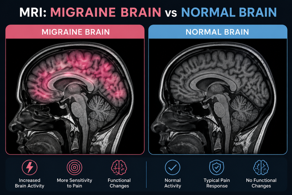
A scan-reading guide to normal MRIs, migraine-associated white-matter spots, and findings that need a different explanation.
A brain MRI can feel like it should provide a simple visual answer to migraine: one scan should look like a migraine brain and another should look normal. In routine medical care, it usually does not work that way.
Migraine is primarily a disorder of brain function and sensory processing. A person can have frequent, disabling migraine attacks and still have a structurally normal MRI. When an MRI does show small bright areas in the white matter, those findings are often nonspecific and can occur in people with migraine, with aging, with vascular risk factors, or in people who have no headache disorder at all.
The practical difference between an MRI migraine brain vs normal brain is therefore not a single signature image. Doctors interpret the scan together with the headache pattern, neurologic examination, age, vascular risks, aura symptoms, and the size, shape, location, and behavior of any MRI findings.
| KEY TAKEAWAYS
· Most people with a typical migraine pattern and a normal neurologic examination do not need brain imaging solely to confirm migraine. · A routine structural MRI can be completely normal even when migraine attacks are severe or frequent. · Some people with migraine have small white matter hyperintensities, often described as tiny bright spots on T2 or FLAIR images. · A 2025 systematic review found that migraine-associated white matter findings are commonly small, supratentorial, and concentrated in deep frontal or parietal white matter, but their clinical significance remains uncertain. · White spots do not automatically mean multiple sclerosis, stroke, dementia, or a brain tumor. The MRI pattern and the clinical history determine what they mean. · Imaging becomes more important when a headache is sudden, new or changing, occurs with a neurologic deficit, starts after age 50, follows trauma, or appears with other red flags. |
Think of the MRI as Three Possible Reports
A useful way to understand migraine imaging is to imagine three common report patterns rather than searching for one “migraine picture.”
| MRI Pattern | What the Radiologist May See | How It Is Usually Interpreted |
| Report A: Structurally normal MRI | No mass, acute stroke, bleeding, hydrocephalus, or significant white matter abnormality | Very common in people with migraine; does not argue against a migraine diagnosis |
| Report B: A few small nonspecific white matter foci | Tiny T2/FLAIR bright spots, often in deep or subcortical white matter | Can be seen with migraine, aging, or vascular factors; interpreted by pattern and clinical context |
| Report C: Atypical or concerning pattern | Mass effect, acute infarct, unusual enhancement, characteristic demyelinating pattern, or other focal abnormality | May point to a diagnosis other than uncomplicated migraine and usually needs targeted follow-up |
What Does a Normal Brain MRI Look Like in Someone With Migraine?
On a routine MRI, a “normal” brain means the radiologist does not see a structural abnormality that explains the symptoms. The brain tissue, ventricles, major fluid spaces, and visualized structures have an expected appearance for the person’s age, and there is no acute infarct, suspicious mass, major bleeding, or other significant finding.
That result is entirely compatible with migraine. MRI is excellent at showing anatomy, but a standard clinical scan is not designed to diagnose the moment-to-moment network activity that produces migraine pain, light sensitivity, nausea, aura, and sensory amplification.
Mayo Clinic explains this distinction clearly in its current migraine diagnosis guidance: migraine can involve abnormal brain function while the underlying structural MRI remains normal.
This is why a normal MRI should not be interpreted as “the headache is not real.” It means that the scan did not identify a structural cause for the symptoms.
What Are the White Spots Sometimes Seen on a Migraine MRI?
The most discussed structural MRI finding in migraine is the white matter hyperintensity, often abbreviated WMH. These are bright-appearing areas on certain MRI sequences, especially T2-weighted and FLAIR images.
A 2023 meta-analysis of 30 studies involving 3,502 people with migraine reported a pooled WMH prevalence of 44%. The pooled estimate was 45% in migraine with aura and 38% in migraine without aura. The studies were highly variable, so those percentages should not be used to predict whether one individual will have lesions.
More recent evidence remains cautious. A 2025 systematic review covering approximately 3,600 participants found that white matter findings were frequently reported in migraine, but the clinical meaning, progression, and relationship with migraine features remained uncertain. Across studies, the lesions were most often small, located above the tentorium, and concentrated in deep frontal or parietal white matter.
In other words, these spots can be associated with migraine, but they are not a migraine fingerprint. They are nonspecific imaging findings.
How Can Migraine White-Matter Spots Differ From Other MRI Lesions?
Radiologists do not judge a white spot by brightness alone. They look at its location, shape, number, size, whether it contacts the cortex or ventricles, whether it involves the corpus callosum, brainstem, or cerebellum, whether it restricts diffusion, and whether it enhances after contrast.
| Possible Explanation | Typical Imaging Clues | Why Context Matters |
| Migraine-associated pattern | Often small and punctate; commonly deep or subcortical frontal/parietal white matter | Usually nonspecific; clinical context is essential |
| Age/small-vessel pattern | Can involve deep and periventricular white matter; burden often increases with age and vascular risk | May prompt review of blood pressure, diabetes, cholesterol, smoking, and vascular health |
| Multiple sclerosis pattern | More likely to include lesions in characteristic periventricular, juxtacortical/cortical, infratentorial, corpus callosal, or spinal-cord locations | Diagnosis requires the overall MRI pattern plus clinical and sometimes laboratory evidence |
| Acute ischemic stroke | Often follows a vascular territory or shows restricted diffusion on appropriate sequences | Requires urgent clinical correlation |
| Tumor or mass | Usually masslike rather than a few scattered tiny WMHs; may cause swelling, mass effect, or enhancement | Needs specialist evaluation based on the exact lesion type |
A July 2026 Mayo Clinic guide to white spots on brain MRI emphasizes that white-matter spots may be linked with aging, small-vessel disease, migraine, stroke, multiple sclerosis, and other conditions. Location is useful, but it is only one part of the interpretation.
Does Migraine With Aura Look Different From Migraine Without Aura?
There is no routine MRI finding that reliably separates every person with aura from every person without aura. Population research suggests white matter lesions may be somewhat more common in migraine with aura, but an individual patient with aura can have a completely normal MRI, and a patient without aura can have small nonspecific spots.
Aura itself is a temporary neurologic phenomenon, most often visual but sometimes sensory or language-related. It is thought to involve cortical spreading depolarization, a wave of altered electrical and chemical activity across the cortex. A standard structural MRI is not a live map of that event.
Research MRI techniques such as functional MRI, perfusion imaging, diffusion methods, spectroscopy, or connectivity analysis can identify group-level differences in migraine studies. Those findings are scientifically important, but they are not currently a routine clinical test that lets a radiologist look at one scan and say, “this is definitely migraine.”
Can an MRI Tell Whether Migraine Has Damaged the Brain?
The presence of white matter spots often raises this fear, but the evidence does not support treating every migraine-associated WMH as brain damage from repeated attacks.
The American Migraine Foundation notes that these lesions are generally not linked to neurological impairment or cognitive decline in typical migraine patients. Research continues because migraine, especially migraine with aura, has associations with vascular risk in some populations, but an incidental small WMH is not the same as a clinical stroke.
The 2025 systematic review reached a similarly cautious conclusion: WMHs are common in migraine, yet their prognostic significance is still unclear, and their presence alone does not justify changing routine migraine management in an otherwise typical presentation.
Why Might a Doctor Order an MRI for a Headache?
The purpose of MRI is usually not to prove that migraine exists. It is to investigate features that make a secondary cause more plausible.
The current American College of Radiology Appropriateness Criteria for Headache state that imaging is usually not appropriate for a typical primary migraine or tension-type headache with a normal neurologic examination and no red flags. Imaging becomes appropriate in several higher-risk scenarios.
- A first or worst headache, especially a sudden thunderclap headache that reaches maximum intensity rapidly.
- A major change in headache frequency, severity, duration, or clinical pattern.
- New weakness, numbness, trouble speaking, persistent visual loss, confusion, seizure, or another focal neurologic deficit.
- New headache beginning after age 50.
- Headache associated with fever, cancer, significant immunocompromise, or recent head trauma.
- Features suggesting abnormal intracranial pressure, such as papilledema or certain positional symptoms.
- New or unusual headache during pregnancy or the postpartum period.
- Unusual, prolonged, or persistent aura, hemiplegic features, brainstem symptoms, or other atypical migraine presentations.
The exact test may be MRI, CT, vascular imaging, or a combination depending on the suspected problem. A sudden thunderclap headache, for example, is approached differently from a slowly changing chronic migraine pattern.
What Does “Nonspecific White Matter Change” Mean on an MRI Report?
The word nonspecific is important. It means the appearance does not point to one unique diagnosis by itself.
A report might say “a few scattered punctate T2/FLAIR hyperintensities in the subcortical white matter, nonspecific.” In a younger person with a long history of migraine and no neurologic deficit, a neurologist may consider the spots compatible with migraine. In an older adult with hypertension, diabetes, smoking history, or high cholesterol, similar spots may be interpreted in the context of small-vessel disease.
If the lesions have a distribution that is more typical of a demyelinating disease, the radiologist may specifically raise that possibility. The next step can include a neurologic examination, comparison with prior scans, spinal MRI, contrast-enhanced imaging, or laboratory testing. The report wording should be read as a description of probability and pattern, not as a diagnosis by itself.
MRI Migraine Brain vs MS Brain: Why the Difference Is Not Just “White Spots”
Migraine and multiple sclerosis can both be associated with white-matter lesions, which is why incidental spots sometimes cause understandable anxiety. But MS is diagnosed from a combination of clinical history and lesions that demonstrate characteristic distribution and, when applicable, dissemination in space and time.
MS lesions more often involve characteristic areas such as periventricular white matter, the cortex or juxtacortical region, the infratentorial brain, the corpus callosum, or spinal cord. Migraine-associated lesions described in research are more commonly small and deep or subcortical, often in the frontal regions.
There can be overlap, and no online checklist can replace a neuroradiologist or neurologist reviewing the actual images. A few nonspecific spots do not establish MS, and a normal brain MRI in a person with typical migraine is common.
What Questions Should You Ask After a Brain MRI?
If the report contains unfamiliar language, these questions are more useful than searching every word individually:
- Was the MRI otherwise normal for my age?
- Are the white-matter spots considered nonspecific, migraine-associated, vascular, or atypical?
- Does the location or shape suggest multiple sclerosis or another demyelinating condition?
- Is there evidence of an old or recent stroke?
- Do I need contrast, vascular imaging, spinal MRI, or another test?
- Should this MRI be compared with an older scan or repeated later?
- Do the findings change my migraine treatment or my vascular-risk management?
A stable scan can also be useful. When prior imaging is available, radiologists can determine whether lesions are unchanged or whether there are genuinely new findings.
When Should a Headache Be Treated as an Emergency?
Seek emergency medical evaluation for a sudden severe headache that reaches peak intensity within seconds to minutes, especially if it is new. Also seek urgent care for headache with new weakness, facial droop, trouble speaking, fainting, severe confusion, seizure, new persistent vision loss, a stiff neck with fever, or symptoms after a significant head injury.
People who are pregnant or recently postpartum, are significantly immunocompromised, have active cancer, or develop a major new headache pattern may also need faster evaluation. The urgency comes from the symptoms and clinical setting, not from whether the person has a previous migraine diagnosis.
Frequently Asked Questions
Can a neurologist diagnose migraine from an MRI?
Usually no. Migraine is primarily diagnosed from symptoms, history, and a neurologic examination. MRI is mainly used when clinicians need to rule out another cause or investigate atypical features.
Does a normal MRI mean I do not have migraine?
No. A structurally normal MRI is common in migraine. Migraine can involve abnormal brain function without a visible structural abnormality on routine imaging.
Are white spots common with migraine?
They can be. Studies report variable rates, and a 2023 meta-analysis estimated a pooled prevalence of about 44% among people with migraine. The spots are nonspecific and are not present in everyone with migraine.
Do migraine white spots turn into multiple sclerosis?
Migraine-associated white matter spots do not simply turn into MS. MS is a separate disease diagnosed from its clinical presentation and characteristic imaging and laboratory evidence.
Can MRI distinguish migraine aura from a stroke?
MRI can help evaluate suspected stroke, but the answer depends on timing, MRI sequences, symptoms, and other tests. A first episode of new weakness, speech trouble, or persistent visual loss should be treated urgently rather than assumed to be aura.
Do I need another MRI every year if I have migraine?
Usually not when migraine symptoms are stable, the neurologic examination is normal, and a prior MRI was reassuring. Repeat imaging is driven by new clinical features, concerning findings, or a specific monitoring plan.
Can a brain tumor be mistaken for migraine?
Some tumors can cause headache, but a tumor usually produces a structural abnormality on imaging rather than the tiny nonspecific white-matter spots commonly discussed in migraine. New neurologic deficits or a major change in headache pattern warrant medical evaluation.
The Bottom Line
When comparing an MRI migraine brain vs normal brain, the most important fact is that the two can look exactly the same on a routine structural scan. Migraine is not diagnosed by finding one characteristic lesion.
Some people with migraine have small white matter hyperintensities, often in deep or subcortical frontal and parietal regions. These findings are usually nonspecific, and current research still does not establish that they represent clinically meaningful brain damage in a typical migraine patient.
What matters most is the pattern. A radiologist considers lesion size, location, shape, enhancement, diffusion, age, vascular risk factors, symptoms, and prior scans. If the headache pattern is typical and the neurologic examination is normal, major imaging abnormalities are uncommon. If the symptoms are new, sudden, changing, or neurologically unusual, imaging can become an important part of ruling out a secondary cause.
Medical note: This article provides general educational information and is not a diagnosis. Brain MRI findings must be interpreted by a qualified clinician or radiologist in the context of symptoms, examination, age, health history, and prior imaging. Evidence and imaging guidance were checked against current U.S. sources available in August 2026.