
Author

admin

Browsing

Glutathione is gaining attention in the health and wellness world, particularly among people interested in antioxidants, nutrition, and healthy aging. But while many people have heard of glutathione supplements, fewer understand how the body naturally produces this important antioxidant.

The human body makes its own glutathione using three amino acids obtained through food and normal metabolic processes. These building blocks, known as glutathione precursors, play an important role in maintaining the body’s natural antioxidant defenses.

Understanding how glutathione is produced, what nutrients support its formation, and how different supplements work can help consumers make more informed wellness decisions.

What Is Glutathione and Why Is It Important?

Glutathione is a naturally occurring antioxidant found in nearly every cell of the human body.

It is made from three amino acids: glutamate, cysteine, and glycine.

One of its primary functions is helping cells manage oxidative stress. This occurs when reactive oxygen species accumulate faster than the body’s protective systems can manage them.

Reactive oxygen species are produced naturally during metabolism. In normal amounts, they serve useful biological functions. However, excessive oxidative activity can damage important cellular structures.

Glutathione helps maintain a healthy balance by participating in antioxidant reactions that protect cells.

It also supports several other biological processes, including normal immune cell function, protein maintenance, and certain reactions involved in processing compounds in the liver.

Because of these functions, researchers continue studying glutathione’s relationship with cellular health and aging.

However, glutathione’s importance does not mean that increasing its levels automatically prevents illness or improves overall health.

What Are Glutathione Precursors?

A precursor is a substance used by the body to produce another compound.

Glutathione precursors are the amino acids needed to manufacture glutathione naturally.

The three primary building blocks are:

  • Glutamate: An amino acid involved in protein metabolism and numerous cellular processes.
  • Cysteine: A sulfur-containing amino acid that plays a particularly important role in glutathione production.
  • Glycine: An amino acid used in protein synthesis and several other metabolic functions.

Cells combine these amino acids through a series of enzyme-controlled reactions.

First, glutamate and cysteine are joined together. Glycine is then added to form the complete glutathione molecule.

The process depends on nutrient availability, enzyme activity, and the body’s metabolic needs.

This explains why adequate nutrition is important for supporting normal glutathione production.

Why Cysteine Is an Important Glutathione Precursor

Cysteine receives considerable attention because its availability can influence glutathione synthesis under certain conditions.

This amino acid contains sulfur, which is important for glutathione’s antioxidant activity.

The body obtains cysteine through dietary protein and can also produce it through metabolic pathways involving other amino acids.

Cysteine is closely related to cystine, a compound formed when two cysteine molecules are joined together.

Certain nutritional products emphasize cystine-rich proteins because they supply amino acid building blocks involved in glutathione synthesis.

However, providing more cysteine or cystine does not guarantee that the body will produce more glutathione in every circumstance.

Glutathione synthesis is carefully regulated, and the effects of supplementation can vary between individuals.

The Roles of Glycine and Glutamate

Although cysteine often receives the most attention, glycine and glutamate are equally necessary components of the glutathione molecule.

Glycine participates in protein synthesis and other metabolic processes. It is found in many protein-rich foods, including meat, fish, dairy products, and legumes.

The body can also produce glycine naturally.

Researchers have investigated whether additional glycine, sometimes combined with cysteine-related nutrients, can influence glutathione production in certain populations.

Some early findings are promising, but additional research is needed to establish meaningful health benefits.

Glutamate, the third building block, is widely involved in normal metabolism.

Because the body generally maintains glutamate through dietary intake and internal production, supplement discussions tend to focus more on cysteine and glycine.

Nevertheless, all three amino acids are essential to the process.

How Food Supports Natural Glutathione Production

One of the most practical ways to support normal glutathione synthesis is by eating a balanced diet containing adequate protein.

When protein is digested, it breaks down into amino acids that the body uses for numerous purposes, including tissue maintenance and the production of important cellular compounds.

Common protein sources include eggs, poultry, fish, dairy products, beans, lentils, tofu, nuts, and seeds.

A varied diet also provides vitamins and minerals that contribute to normal antioxidant activity.

For example, selenium is required for the activity of certain glutathione-related enzymes, while vitamin C participates in other antioxidant processes.

Vegetables such as broccoli, cabbage, and Brussels sprouts contain naturally occurring compounds studied for their effects on cellular defense systems.

However, no single food guarantees increased glutathione levels.

Overall dietary quality matters more than relying on one particular ingredient.

Understanding Whey Protein and Glutathione Precursors

Whey protein is a complete protein derived from milk. It contains all nine essential amino acids and is widely used to support general nutrition and muscle maintenance.

Certain whey proteins also provide cysteine-containing compounds involved in glutathione production.

This has led to interest in specialized whey-derived nutritional products.

One example is Immunocal, a cystine-rich whey protein product designed to supply amino acid precursors used in the body’s natural glutathione synthesis pathways.

Unlike supplements containing glutathione directly, Immunocal provides protein-derived building blocks that the body can use in its own metabolic processes.

It is important to recognize that not all whey protein products have identical compositions or supporting research.

Readers interested in learning more about Immunocal, its ingredients, and how cystine-rich whey proteins differ from conventional protein supplements can explore the educational information atImmuneGluta.com.

As with any nutritional supplement, consumers should distinguish established nutritional functions from broader health claims that require additional scientific evidence.

Direct Glutathione Supplements Versus Glutathione Precursors

When researching glutathione supplements, consumers generally encounter two different approaches.

The first involves taking products that contain glutathione itself. These supplements may be available as capsules, powders, liquids, or liposomal formulations.

The second approach involves supplying the body with nutrients used to produce glutathione naturally.

These may include cysteine-related compounds, glycine, or specialized protein formulations.

Both approaches have been investigated in scientific studies, but they are not interchangeable.

Some research has reported changes in glutathione measurements following particular interventions. However, results vary depending on the ingredients, dosage, duration, and people studied.

An increase in glutathione levels does not automatically establish improvements in energy, immune health, or longevity.

Understanding these differences is important before choosing a product.

Want to learn more about glutathione and how your body produces it?

Check out our Glutathione Guide for additional information about glutathione, its amino acid precursors, antioxidant health, and the different approaches to supporting natural glutathione production.

Learning how these processes work can make it easier to compare ingredients, understand supplement labels, and recognize exaggerated marketing claims.

What Is N-Acetylcysteine?

N-acetylcysteine, commonly known as NAC, is another compound frequently discussed in connection with glutathione.

NAC can supply cysteine, one of the amino acids required for glutathione synthesis.

It has established medical applications, including treatment of acetaminophen overdose under professional medical supervision.

NAC is also marketed as a dietary supplement and has been studied for other potential applications.

However, its medical uses should not be confused with general wellness claims.

NAC may cause side effects or interact with certain medications. Anyone considering it for a specific health concern should seek qualified medical guidance.

Does Glutathione Production Change With Age?

Researchers have investigated whether glutathione metabolism changes as people get older.

Some studies have found differences in glutathione concentrations or antioxidant balance in certain tissues and populations of older adults.

These findings have contributed to growing interest in glutathione precursors and healthy aging.

However, aging affects individuals differently, and glutathione measurements alone do not provide a complete picture of health.

Researchers are still investigating whether particular supplements produce meaningful improvements in physical function, quality of life, or other outcomes.

There is currently insufficient evidence to claim that glutathione supplements or their precursors reverse aging.

A broader approach that includes nutrition, exercise, sleep, and preventive healthcare remains essential.

How to Choose Glutathione-Related Supplements

With so many products marketed for antioxidant support, understanding supplement labels is important.

Consumers should first identify whether a product contains glutathione directly or provides precursors involved in its synthesis.

The ingredient list should clearly explain what the product contains and how much is included in each serving.

Other considerations include manufacturer transparency, quality-control practices, and whether independent testing is available.

Research involving an individual ingredient does not necessarily prove that every finished product containing that ingredient will produce the same effects.

Consumers should also be cautious about products promising dramatic anti-aging benefits or guaranteed improvements in health.

Supplements may be appropriate in certain circumstances, but they should not replace balanced nutrition or necessary medical treatment.

Everyday Habits That Support Antioxidant Health

Supporting the body’s antioxidant defenses involves more than choosing a supplement.

Eating adequate protein helps supply amino acids needed for normal cellular processes.

Fruits and vegetables provide vitamins, minerals, and plant compounds that contribute to overall nutritional health.

Regular physical activity can help the body maintain healthy metabolic and antioxidant responses.

Adequate sleep supports recovery and normal physiological regulation.

Avoiding tobacco and limiting excessive alcohol consumption can also reduce preventable sources of cellular stress.

These habits have broader evidence supporting their value for long-term health than many products promoted as antioxidant shortcuts.

Final Thoughts

Glutathione is an important antioxidant that the body naturally produces using three amino acids: glutamate, cysteine, and glycine.

These glutathione precursors help explain the connection between dietary protein, cellular metabolism, and antioxidant function.

Specialized nutritional products may supply glutathione directly or provide building blocks involved in its natural production.

Understanding the differences between these approaches allows consumers to evaluate ingredients and supplement claims more carefully.

For most people, the foundation of healthy glutathione metabolism begins with balanced nutrition, adequate protein, regular physical activity, and other established wellness habits.

Learning how glutathione works is a useful first step toward making informed decisions about antioxidant health.


As regenerative medicine has grown rapidly as a field, patients researching treatment options face a genuinely difficult challenge: distinguishing credible, ethical providers from those making unsupported or exaggerated claims about what their specific treatments can realistically achieve for any given patient. This challenge exists globally and deserves serious, careful attention from anyone considering this significant medical decision.

Laboratory accreditation stands as one of the most fundamental factors to verify before committing to any provider whatsoever. A credible clinic should be able to clearly explain exactly where and how cells are processed, under what specific accreditation standards, and whether the facility undergoes regular inspection by an appropriate regulatory body with genuine oversight authority. A provider who cannot answer these questions clearly is signalling something important about their overall approach to patient care and transparency.

Before choosing any provider, patients should specifically ask who reviews their case before treatment is confirmed to proceed – genuine, ethical practice involves more than a single specialist making this determination in isolation from other qualified input. How a provider communicates about expected outcomes often provides the clearest window into their overall trustworthiness as a medical practice. Be genuinely cautious of any provider who guarantees a cure or promises a specific fixed percentage of improvement without appropriate caveats about individual variation.

Reputable stem cell treatment centers are generally willing to discuss laboratory accreditation, treatment evaluation processes, and realistic outcome expectations openly and in genuine detail, rather than deflecting this kind of reasonable scrutiny from prospective patients. Aftercare planning deserves equal attention during this evaluation process – ask specifically what monitoring is planned following any procedure and how follow-up care will be coordinated if you’re travelling internationally and returning home afterward.

Patients are encouraged to review a provider’s approach to case evaluation and aftercare as a genuinely key indicator of overall treatment quality and trustworthiness, since these operational details often reveal considerably more about a clinic’s actual standards than any marketing materials or promotional claims could ever convey to a prospective patient making this significant decision.

If you are wondering what are kcals, the simplest explanation is that if an app says 500 kcal and a U.S. food label says 500 Calories, they are telling you the same thing.

The word kcal can make nutrition look more technical than it really is. In everyday food and fitness language, one kilocalorie, written as 1 kcal, is the same amount of energy Americans usually call 1 Calorie with a capital C.

So a meal listed as 650 kcal contains the same food energy as a meal listed as 650 Calories on a U.S. Nutrition Facts label. You do not multiply the food-label number by 1,000. The 1,000-to-1 difference matters only when you compare a kilocalorie with the much smaller scientific unit called a calorie with a lowercase c.

That distinction explains why nutrition apps, international food packages, exercise machines, and U.S. labels can use different-looking terms while reporting the same practical energy amount.

KEY TAKEAWAYS

·        1 kcal equals 1 food Calorie, also written as 1 Cal with a capital C.

·        1 kcal equals 1,000 small scientific calories.

·        A U.S. Nutrition Facts label that says 200 Calories is effectively reporting 200 kcal.

·        Carbohydrate and protein generally provide about 4 kcal per gram, while fat provides about 9 kcal per gram.

·        The FDA uses 2,000 Calories per day as a general label-reference amount, not as a personalized prescription.

·        Your actual energy needs vary with age, body size, activity, life stage, health, and other individual factors.

The 10-Second Decoder: kcal, Calorie, and calorie

Term Symbol Where You See It What It Means
1 calorie cal Small scientific unit Energy needed to raise about 1 gram of water by 1°C
1 kilocalorie kcal Food-energy unit 1,000 small calories
1 Calorie Cal U.S. nutrition term The same energy as 1 kcal

In normal nutrition conversation, people usually write ‘calories’ with a lowercase c even when they technically mean kilocalories. That shortcut is so common that it rarely causes a practical problem. The confusion appears mostly when someone notices kcal on an app or international label and assumes it must be a different number.

Why Does a Kilocalorie Equal 1,000 Calories?

The prefix kilo means 1,000. A kilometer is 1,000 meters, and a kilogram is 1,000 grams. The same logic applies to energy units: one kilocalorie is 1,000 calories.

A small calorie is traditionally defined in terms of the energy required to raise the temperature of a small amount of water. A kilocalorie scales that energy up by 1,000. In modern scientific work, energy is usually expressed in joules rather than calories.

The conversion is straightforward: 1 small calorie equals 4.184 joules, so 1 kilocalorie equals 4,184 joules, or 4.184 kilojoules.

That is why labels in some countries show both kcal and kJ. For example, 250 kcal is about 1,046 kJ.

Why Do U.S. Food Labels Say Calories Instead of kcal?

The FDA Nutrition Facts Label guidance uses the word Calories for the energy in a serving of food or beverage. In this context, the number is a kilocalorie value even though the label does not print kcal next to it.

If a cereal label says 140 Calories per serving, that means 140 kcal. If a European nutrition app shows the same serving as 140 kcal, the energy amount has not changed.

The FDA also emphasizes serving size because calorie information is usually listed per serving. If a package contains two servings and you eat the whole package, you generally consume twice the Calories shown for one serving.

Where Do Kcals in Food Come From?

Food energy comes mainly from carbohydrate, protein, and fat. Alcohol also contributes energy.

For ordinary nutrition calculations, the familiar Atwater factors are:

Macronutrient Approximate Energy
Carbohydrate About 4 kcal per gram
Protein About 4 kcal per gram
Fat About 9 kcal per gram

The USDA’s FoodData Central reports food energy in kcal and explains that many energy values are calculated using Atwater factors. This is one reason a food’s calorie total can often be estimated from its grams of carbohydrate, fat, and protein.

For example, imagine a food containing 20 grams of carbohydrate, 10 grams of protein, and 5 grams of fat. A rough calculation would be 80 kcal from carbohydrate, 40 kcal from protein, and 45 kcal from fat, for about 165 kcal total.

Real labels can differ slightly from a simple calculation because food composition, fiber, sugar alcohols, specialized factors, laboratory methods, and FDA rounding rules can affect the displayed number.

If My Fitness Watch Says I Burned 400 Calories, Is That 400 kcal?

Yes. In everyday fitness language, a device reporting 400 calories burned is effectively reporting about 400 kcal of energy expenditure.

The harder question is whether the estimate is accurate. Watches and exercise machines infer calorie burn from inputs such as heart rate, movement, age, body size, workout type, and proprietary algorithms. They do not directly measure every calorie your body uses.

Treat workout calorie numbers as estimates rather than permission to replace every displayed calorie with extra food. Two devices can produce different numbers for the same session, and exercise energy expenditure varies between people.

Does 2,000 kcal Mean Everyone Should Eat 2,000 Calories?

No. The 2,000-Calorie figure on U.S. labels is a reference amount used to make nutrition information easier to compare.

The FDA notes that an individual’s needs can be higher or lower depending on factors including age, sex, height, weight, and physical activity. Pregnancy, breastfeeding, growth, illness, training volume, and weight-management goals can also change energy needs.

The current U.S. Dietary Guidelines for Americans, 2025-2030, were released in 2026. They emphasize overall eating patterns and nutrient-rich food choices rather than treating one calorie level as correct for everyone.

For current federal nutrition guidance, see the Dietary Guidelines for Americans. Personalized calorie targets are better estimated from individual characteristics than copied from the 2,000-Calorie label reference.

Why Two Foods With the Same Kcals Can Affect You Differently

Calories measure energy, not overall nutritional quality.

A 250-kcal snack could be a combination of fruit, yogurt, and nuts, or it could be a sugary beverage and candy. The energy total may be similar, but the foods can differ greatly in protein, fiber, vitamins, minerals, added sugars, saturated fat, volume, and how filling they feel.

Protein and fiber often contribute to fullness. Foods with water and fiber can provide more volume for the same calorie total. Highly energy-dense foods can pack many kcal into a small portion.

This is why calorie awareness can be useful without making calories the only thing you consider. A better label-reading approach is to look at serving size, Calories, protein, fiber, added sugars, saturated fat, sodium, and the ingredient list together.

Kcal vs kJ: What If the Label Uses Kilojoules?

Kilojoules measure the same thing, energy, using the international SI system.

A useful conversion is:

1 kcal = 4.184 kJ

So 100 kcal is about 418 kJ, and 500 kcal is about 2,092 kJ.

You usually do not need to convert manually. Many labels outside the United States list both values, and nutrition apps often let you choose a preferred energy unit.

Common Kcal Mistakes to Avoid

Mistake 1: Multiplying Food-Label Calories by 1,000

If a U.S. label says 300 Calories, it already means about 300 kcal. You should not interpret it as 300 small scientific calories or convert it to 300,000 food Calories.

Mistake 2: Treating the Serving Size as a Recommendation

Serving size tells you the amount used for the nutrition calculation. The FDA specifically notes that serving size reflects how much people typically consume, not how much an individual should necessarily eat.

Mistake 3: Assuming Low-Calorie Automatically Means Nutritious

A low-kcal item can still be high in sodium or low in fiber, protein, vitamins, and minerals. Likewise, calorie-dense foods such as nuts can provide useful nutrients. Context and portion size matter.

Mistake 4: Treating Exercise Calories as Exact

Wearable and machine estimates are useful for trends, but they are not laboratory measurements. Avoid building a precise eating plan around one workout number.

Mistake 5: Using a Fixed Calorie Formula for Weight Change

Weight change is influenced by energy intake and expenditure, but the body adapts over time. Metabolism, body composition, water balance, activity, appetite, and adherence all affect real-world results.

Simple rules such as assuming a fixed calorie deficit will always produce the same amount of weight loss can be useful as rough illustrations, but they should not be treated as precise biological predictions.

How Should You Use Kcals When Reading a Nutrition Label?

A simple five-step scan works well:

  • Check the serving size first.
  • Look at Calories, which are equivalent to kcal.
  • Decide how many servings you are actually eating.
  • Check protein, fiber, added sugars, saturated fat, and sodium.
  • Compare similar foods using the same serving amount whenever possible.

Suppose a package lists 220 Calories per serving and contains three servings. Eating one serving gives you 220 kcal. Eating half the package gives you about 330 kcal. Eating the whole package gives you 660 kcal.

That simple serving-size check prevents more confusion than almost any advanced calorie calculation.

Frequently Asked Questions

Is 1 kcal the same as 1 Calorie?

Yes. In nutrition, 1 kcal equals 1 Calorie with a capital C. This is the unit used for food energy on U.S. labels, even though labels usually print the word Calories rather than kcal.

Is 100 kcal the same as 100 calories on a food label?

Yes. A U.S. food label showing 100 Calories is reporting approximately the same amount of food energy as 100 kcal.

How many small calories are in 1 kcal?

There are 1,000 small scientific calories in 1 kilocalorie.

How many joules are in 1 kcal?

One kilocalorie equals 4,184 joules, or 4.184 kilojoules.

Do kcal make you gain weight?

Kcal are simply a measure of energy. Consistently consuming more energy than your body uses can contribute to weight gain, while consistently consuming less can contribute to weight loss. Real body-weight changes are affected by many factors and are not perfectly predicted by one fixed formula.

Why does Europe use kcal?

Many food-labeling systems outside the United States display energy as kcal, often alongside kJ. U.S. labels usually use Calories, but the practical food-energy unit is the same.

Should I count kcal every day?

Not everyone needs to. Tracking can be useful for some goals, but meal quality, hunger and fullness, medical needs, activity, and sustainability matter too. People with specific nutrition or health concerns may benefit from individualized guidance from a registered dietitian or clinician.

The Bottom Line

Kcals are simply units of food energy. One kcal equals one food Calorie, so the numbers you see in a nutrition app, on an international food package, or on a U.S. Nutrition Facts label can usually be compared directly.

The technical difference only appears when you compare a kilocalorie with a small scientific calorie: 1 kcal equals 1,000 calories. For everyday eating, that distinction is mostly terminology.

The more useful skill is knowing what the number can and cannot tell you. Calories help describe energy intake, but they do not measure overall diet quality. Start with serving size, understand that Calories and kcal are equivalent in nutrition, and use the rest of the label to judge the food’s nutrient profile.

This article is for general educational purposes and is not individualized nutrition or medical advice. Nutrition guidance and labeling information were checked against current U.S. federal sources available in August 2026.

A 2026 visual guide to borders, center clearing, scaling, color on different skin tones, nummular eczema, contagiousness, testing, and treatment mistakes

When comparing ringworm vs eczema pictures, the two conditions can look surprisingly similar. Both can cause round, itchy, scaly patches, and the confusion becomes even greater when the eczema is nummular eczema, a type that forms coin-shaped spots.

The most useful visual clue is often the edge of the rash. Ringworm tends to have a more active, raised, scaly outer border with relatively clearer skin toward the center, while eczema is often more uniformly inflamed, dry, cracked, or weepy across the patch.

That rule is helpful, but it is not reliable enough to diagnose every rash from a photo. Ringworm may lose its classic ring shape on certain body sites, eczema can clear in the center as it heals, and steroid cream can change the appearance of a fungal infection. Skin tone also changes how redness appears. On darker skin, both conditions may look brown, gray, purple, or darker than the surrounding skin rather than bright red.

The fastest visual distinction

Ringworm often expands outward with a sharper, raised, scaly edge and partial clearing in the center. Nummular eczema is more likely to look like a solid coin-shaped patch that is dry, crusty, cracked, or weepy throughout. If the picture does not clearly fit either pattern, a clinician can examine the skin and, when needed, test a scraping for fungus.

Key Takeaways

  • Ringworm is a contagious fungal infection. Eczema is inflammatory and not contagious.
  • Ringworm often has a raised, scaly outer border with relative clearing in the center.
  • Nummular eczema is the eczema type most likely to mimic ringworm because its patches are round or oval.
  • Eczema is often itchy and dry, cracked, crusted, blistered, or oozing across the patch.
  • On darker skin, both rashes may look brown, gray, purple, or darker rather than bright red.
  • Ringworm can spread to other people, animals, and body sites. Eczema cannot be caught from someone else.
  • Do not use steroid cream alone on a rash that could be ringworm. Steroids can worsen or disguise fungal infection.
  • A clinician can use a skin scraping or fungal test when the appearance is unclear.

Visual Snapshot: Ringworm vs. Eczema in Pictures

Picture Clue Ringworm Eczema / Nummular Eczema
Shape Often round or ring-shaped and gradually enlarging Round/oval in nummular eczema, or irregular in other eczema types
Outer edge Often raised, scaly, and more active than the center Usually less sharply defined; may be dry, crusted, cracked, or inflamed throughout
Center May look clearer or closer to normal skin May stay inflamed; can also flatten or clear as a patch heals
Scale Often concentrated near the advancing border Can cover much or all of the patch
Moisture Usually dry/scaly on the body May ooze, blister, crust, or become very dry
Number of spots May start as one patch and spread or form overlapping rings May appear as multiple coin-shaped spots, often on limbs
Contagious? Yes No

For medically reviewed examples of the classic fungal patterns, see the American Academy of Dermatology ringworm photo guide.

What Does Ringworm Look Like in a Picture?

Ringworm of the body, called tinea corporis, usually starts as a small scaly patch and slowly expands. The growing edge often looks more inflamed than the middle. That outer rim may be slightly raised, bumpy, or flaky, giving the rash the ring-like appearance that produced the name.

  • A round or oval patch that gradually becomes larger.
  • A raised or wavy border that looks more active than the center.
  • Scale or small bumps concentrated near the edge.
  • Partial clearing in the middle, although this is not always present.
  • One ring or several rings that can overlap.
  • Itching that ranges from mild to intense.

The CDC notes that ringworm on most body skin may have a clear or scaly center, a slightly raised border, and red, red-purple, brown, gray, or black discoloration depending on skin tone. Scalp, groin, foot, beard, palm, and nail infections can look very different and may not form a clean ring.

The CDC ringworm signs and symptoms page includes updated 2026 descriptions for ringworm on the skin, scalp, feet, groin, beard, and nails.

What Does Eczema Look Like in a Picture?

Eczema is a group of inflammatory skin conditions rather than one single rash pattern. Atopic dermatitis, the most common form, often causes very itchy, dry, scaly skin that can become thickened, cracked, raw, blistered, or crusted after scratching.

A typical eczema picture may show patches with softer or less distinct borders than ringworm. The skin may look rough or leathery from repeated rubbing, and the rash may appear in several areas associated with age and body site, such as the face in babies, elbow and knee creases in many children, or hands, neck, eyelids, and flexural areas in adults.

  • Dry, rough, scaly skin around and between patches.
  • Intense itch, often severe enough to disturb sleep.
  • Cracks, scratch marks, or thickened skin from repeated rubbing.
  • Small bumps, blisters, leaking fluid, or crusting during some flares.
  • Patches that return in the same areas over time.
  • Color that may be pink/red on lighter skin or purple, gray, brown, or darker/lighter than surrounding skin on darker tones.

Why Nummular Eczema Looks So Much Like Ringworm

Nummular eczema, also called nummular dermatitis or discoid eczema, is the main reason a search for ringworm vs. eczema pictures can be confusing. It forms round or oval spots that resemble coins, sometimes with enough scaling and center change to imitate a fungal ring.

The American Academy of Dermatology describes nummular eczema as often beginning with tiny bumps or blister-like sores that crust and merge into a raised coin-shaped patch. The patches frequently develop on the legs, forearms, or backs of the hands and can appear on both sides of the body.

Clue More Suggestive of Ringworm More Suggestive of Nummular Eczema
Border Sharper, raised, expanding, scaly rim Less consistently ring-like; may be inflamed across the whole coin
Center Often clearer than the edge May remain dry/scaly, ooze, crust, or later flatten while healing
Skin between patches May look normal Often very dry
Typical sites Anywhere on skin, plus site-specific tinea patterns Often legs, forearms, backs of hands
Trigger pattern Exposure to infected person, pet, surface, or body site Dry skin; may follow a cut, scrape, burn, or insect bite
Spread to others Possible No

For photographs specifically showing coin-shaped eczema, see the AAD nummular eczema signs and symptoms gallery.

How Do the Rashes Look on Darker Skin Tones?

Color words such as red and pink can be misleading when comparing dermatology pictures. In brown and Black skin, inflammation may appear deep brown, gray, violet, purple, or darker than the surrounding skin. Some healing eczema patches can also become temporarily lighter or darker than the person’s baseline skin color.

Ringworm can also be less obviously red on darker skin. The CDC describes patches as potentially red-purple, brown, gray, or black, while the AAD notes that ringworm on skin of color is often brown or gray. This is why shape, scale, border activity, itching pattern, location, and spread are usually more useful than color alone.

Picture-search warning

Online image results often overrepresent lighter skin. A rash does not have to be bright red to be active ringworm or eczema. If the shape seems suspicious but the color looks different from the pictures you found, do not rule either condition out on color alone.

Where on the Body Can Location Help?

Location can provide a clue, but it cannot diagnose the rash by itself. Ringworm changes its appearance depending on the body site, while different types of eczema also favor certain locations.

Body Area Ringworm Pattern Eczema Pattern
Arms / trunk Classic ring-shaped tinea corporis common here Atopic or nummular eczema can also affect these areas
Legs Possible Very common site for nummular eczema
Elbow / knee creases Possible Common pattern in atopic dermatitis
Scalp Scaly patch, broken hairs or hair loss; often needs oral antifungal treatment Dry, itchy or inflamed scalp possible with eczema types
Feet Athlete’s foot: peeling, scaling, cracking, especially between toes or soles Eczema can affect feet but usually does not follow classic athlete’s-foot pattern
Groin Jock itch, often with a scaly advancing edge Contact or atopic dermatitis may occur but is treated differently

Is Ringworm Contagious While Eczema Is Not?

Yes. Ringworm is caused by dermatophyte fungi and can spread through direct skin contact, infected animals, contaminated objects, and shared surfaces. Athlete’s foot, jock itch, scalp ringworm, and many fungal nail infections are related tinea infections caused by the same general group of fungi.

Eczema is not an infection and cannot be caught from another person. A person with eczema can develop a secondary bacterial, viral, or fungal infection on damaged skin, but the underlying eczema itself is not contagious.

How Can a Doctor Tell Ringworm From Eczema?

Many cases can be diagnosed by appearance and history, but circular rashes are one of the situations where testing may be useful. A clinician may gently scrape scale from the active edge of a suspected fungal rash and examine or send the sample for fungal testing. In selected cases, culture, dermoscopy, a swab for infection, or a skin biopsy may be considered.

Testing becomes especially useful when a rash has been treated but is not improving, when it has an unusual appearance, when the scalp or nails are involved, or when a person has already applied a topical steroid that may have changed the fungal rash.

The Treatment Mistake That Can Make the Pictures Harder to Read

A common mistake is applying an over-the-counter steroid cream to any itchy rash. Topical corticosteroids are useful medications for many forms of eczema because they reduce inflammation and itching. They are not antifungal treatment.

The CDC specifically warns against using steroid creams or ointments for ringworm or a rash that may be ringworm. A steroid can suppress redness and scaling without killing the fungus, allowing the infection to spread while looking less typical. This altered appearance is sometimes called tinea incognito.

If you are unsure which rash it is

Avoid treating an undiagnosed circular rash with steroid cream alone. Ringworm usually needs antifungal treatment, while eczema often needs skin-barrier care and anti-inflammatory treatment. Using the wrong treatment can make the rash harder to recognize and delay improvement.

How Is Ringworm Usually Treated?

Ringworm treatment depends on the body site. Limited tinea corporis on ordinary skin is often treated with an antifungal cream, lotion, powder, or ointment. Scalp ringworm usually requires prescription oral antifungal medication because topical creams do not reach infected hair follicles adequately.

Treatment should continue for the recommended duration even if the rash starts looking better sooner. Clothes, towels, bedding, sports gear, and household contact with infected pets may also need attention to reduce reinfection or spread.

How Is Eczema Usually Treated?

Eczema treatment depends on the type and severity. For nummular eczema, dermatologists commonly emphasize restoring the skin barrier with regular fragrance-free cream or ointment moisturizers and avoiding harsh soaps, hot water, and other irritants.

Prescription or clinician-directed anti-inflammatory treatment may include topical corticosteroids or nonsteroid medicines. More extensive or persistent eczema can require additional therapies. If the skin becomes infected, that infection may need separate treatment.

When Should You Stop Comparing Pictures and Get Medical Care?

  • The rash is on the scalp, beard area, nails, around the eyes, or a large body area.
  • There is hair loss, a painful swollen scalp area, pus, or significant crusting.
  • The rash is rapidly spreading or appears after close contact with a known ringworm infection.
  • A circular rash keeps returning or does not improve with appropriate over-the-counter antifungal treatment.
  • You used steroid cream and the rash became larger, less typical, or more widespread.
  • The skin is painful, warm, swollen, draining pus, or covered with yellow/golden crusts.
  • You have fever or feel ill along with the rash.
  • The person with the rash is an infant, is immunocompromised, or has a condition that increases infection risk.

Frequently Asked Questions

What is the easiest way to tell ringworm from eczema in a picture?

Look at the outer border and the center. Ringworm often has a raised, scaly, active edge with relatively clearer skin toward the center. Eczema is more likely to be dry, cracked, crusted, or inflamed throughout the patch. However, nummular eczema can mimic ringworm closely, so this clue is not enough for every case.

Can eczema form a perfect circle?

Yes. Nummular eczema can form round or oval coin-shaped patches and may be mistaken for ringworm. Some patches can also clear in the center as they heal.

Can ringworm look like eczema?

Yes. Ringworm can be dry, itchy, scaly, and inflamed, especially before a clear ring develops. Steroid treatment can also alter the appearance and make the fungal rash resemble eczema or another dermatitis.

Is ringworm always a red circle?

No. It may be pink or red on lighter skin but brown, gray, red-purple, or darker on other skin tones. Ringworm on the feet, scalp, groin, palms, beard, or nails may not look like a classic red ring at all.

Is eczema contagious?

No. Eczema is not contagious. Ringworm is contagious because it is caused by fungi that can spread between people, animals, and contaminated objects or surfaces.

Can I put hydrocortisone on a rash if I am not sure?

It is better not to use steroid cream alone on an undiagnosed rash that could be ringworm. The CDC warns that steroids can make ringworm worse and can change its appearance. If the diagnosis is uncertain, ask a clinician or pharmacist about the safest next step.

How long does ringworm take to appear after exposure?

The CDC says symptoms of ringworm generally appear about 4 to 14 days after skin contact with the fungi that cause the infection.

Can a doctor test for ringworm?

Yes. Depending on the site and appearance, a clinician can take a small skin scraping, hair sample, or nail sample for fungal testing. Testing can be especially helpful when ringworm and eczema look alike.

Bottom Line: Pictures Help, but the Border Is Only a Clue

Ringworm vs. eczema pictures are useful because they teach you where to look: the edge, the center, the scale pattern, the body location, and whether the rash is spreading. A classic ringworm patch grows outward with a more active scaly rim and some center clearing. Classic eczema looks more diffusely dry, itchy, cracked, inflamed, or weepy.

The difficult cases are usually nummular eczema and steroid-altered ringworm. Both can create round patches that break the usual visual rules. That is why a recurring circular rash should not be treated based on internet photos alone, especially when a steroid or antifungal has already been tried without success.

If you are unsure, the most useful next step is not finding a closer-looking photo. It is getting the rash examined and, when appropriate, tested. That matters because ringworm requires antifungal treatment while eczema is treated by calming inflammation and repairing the skin barrier.

Medical note: This article is for general education and cannot diagnose a rash from a description or photograph. It was updated for U.S. readers using information available in August 2026 from the CDC, American Academy of Dermatology, National Eczema Association, and the supplied HealthCentral reference. Seek professional care for uncertain, severe, spreading, infected, scalp, nail, or treatment-resistant rashes.

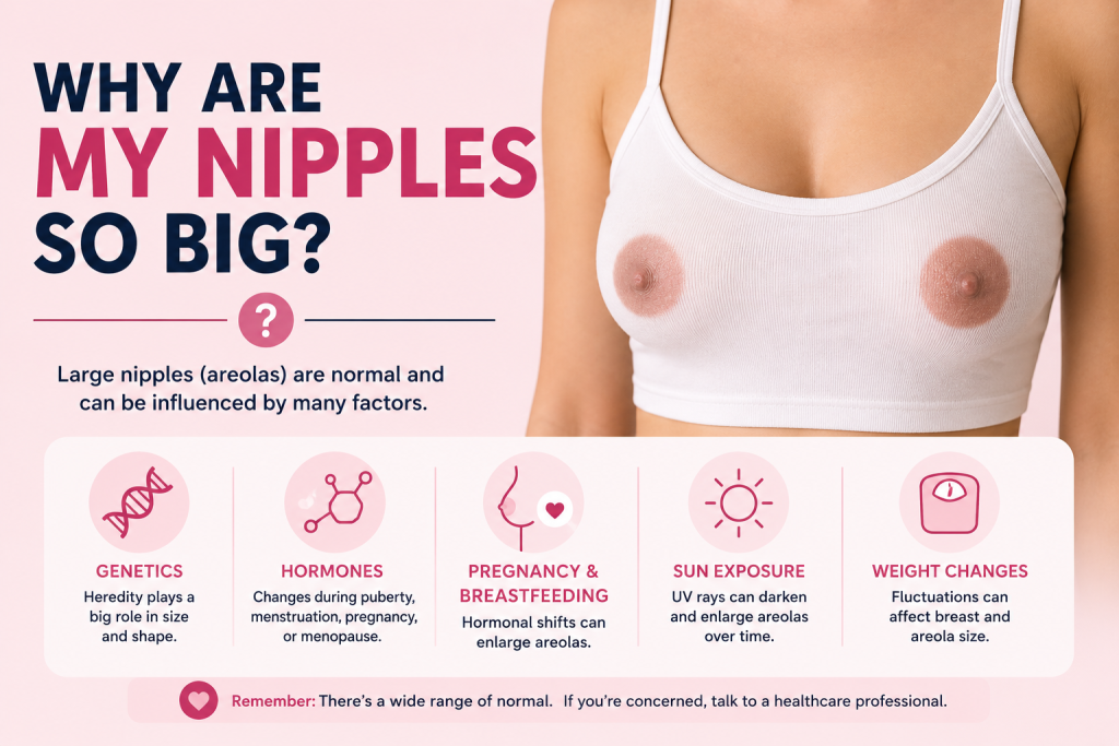
Nipples and areolas vary widely, so if you are asking why are my nipples so big, the most useful clues are whether the appearance is normal for you, whether it changed recently, and whether other breast symptoms appeared at the same time.

The first step is to identify which part looks larger. The nipple is the raised or flat structure in the center of the breast. The areola is the circular area of darker or differently colored skin surrounding it. Many people who are concerned about “large nipples” are actually noticing large areolas.

Both nipple and areola size vary naturally from person to person. Genetics, breast size, puberty, hormonal changes, pregnancy, breastfeeding, weight changes, aging, and benign breast conditions can all affect how the nipple-areola complex looks. One side can also be slightly different from the other without signaling a health problem.

Cleveland Clinic notes in its nipple anatomy guide that nipple appearance varies from person to person. Nipples can protrude, lie flat, or point inward, and the surrounding areola is a separate structure whose size and color also vary widely.

KEY TAKEAWAYS

·        There is no single “correct” nipple or areola size. Normal anatomy has a broad range.

·        If the nipple-areola area has always looked large and both sides are otherwise healthy, it is usually normal variation.

·        Puberty, pregnancy, breastfeeding, menstrual hormones, weight changes, and breast growth can make nipples or areolas look larger.

·        In males, gynecomastia or increased chest fat can make the nipple and areola look wider or puffier.

·        A recent one-sided change deserves more attention than a stable lifelong difference.

·        New nipple inversion, bloody or spontaneous discharge, a lump, skin dimpling, persistent scaling, or redness should be evaluated by a healthcare professional.

First, Are You Looking at the Nipple or the Areola?

Part What It Is
Nipple The central structure that may protrude, lie flat, or turn inward. It contains small openings for milk ducts.
Areola The circular pigmented skin around the nipple. It can be small, large, round, oval, light, dark, smooth, or bumpy.
Montgomery glands Small oil-producing glands in the areola that can look like tiny bumps and may become more noticeable with hormonal changes.

This distinction matters because an areola can expand as the breast grows even when the central nipple has barely changed. Darker pigmentation can also make an areola seem visually larger than it is.

Is There a Normal Nipple Size?

There is a wide normal range rather than one ideal measurement. Cleveland Clinic reports an average nipple width of about 10 to 12 millimeters, but an individual nipple can be smaller or larger and still be healthy. Appearance also changes with temperature, touch, hormonal shifts, and sexual arousal because smooth muscle in the nipple can contract.

Areola size varies even more. People with larger breasts often have larger areolas, but breast size does not perfectly predict areola size. Two healthy people of the same age and body size can have very different nipple and areola proportions.

Symmetry is also not required. One breast, nipple, or areola may be somewhat larger than the other. A stable difference that has been present for years is generally less concerning than a new change.

Why Can Nipples or Areolas Look Bigger Over Time?

Puberty and Normal Breast Development

During puberty, rising hormones stimulate breast tissue and the duct system to grow. Johns Hopkins Medicine notes that the areola becomes larger as breast development progresses. The two breasts do not always develop at exactly the same speed, so temporary asymmetry is common.

Pregnancy and Breastfeeding

Pregnancy can cause some of the most noticeable changes. The breasts enlarge, areolas often become wider and darker, nipples may become more prominent, and Montgomery glands can become easier to see. These changes help prepare the breast for feeding.

After pregnancy and breastfeeding, the nipple-areola area may move back toward its previous appearance, but it does not always return to exactly the same size or color. Persistent changes after pregnancy can still be normal.

Menstrual Hormone Changes

Hormonal shifts during the menstrual cycle can make breasts feel fuller, swollen, or tender. The change is usually temporary and affects both breasts. Some people notice that nipples feel more sensitive or look more prominent before a period even if the actual tissue size has changed very little.

Weight Gain or Weight Loss

Breasts contain a substantial amount of fatty tissue. When breast volume increases with weight gain, the skin and areola may stretch and look larger. Weight loss can make the areola appear relatively different as breast volume changes, although the skin does not always shrink in perfect proportion.

Genetics and Natural Proportions

Some people simply have larger nipples or areolas from adolescence onward. Genetics influence breast size, skin pigmentation, tissue distribution, and the proportions of the nipple-areola complex. If your appearance has been stable and there are no other symptoms, size alone is not usually a sign of disease.

Montgomery Glands and Areola Bumps

The small bumps on the areola are often Montgomery glands, not abnormal growths. Cleveland Clinic explains that these glands release oil that protects the nipple and areola. They can become more noticeable during pregnancy and breastfeeding, but their number and visibility vary even outside those life stages.

A single gland can occasionally become blocked or inflamed. A tender red bump that behaves like a pimple may need simple local care or medical evaluation if it persists, worsens, or appears infected.

What If You Are Male and the Nipple Area Looks Larger or Puffy?

In boys and men, a larger or puffier nipple-areola area can be related to gynecomastia, which is an increase in glandular breast tissue. It is common during puberty and can also occur in adulthood when the balance between testosterone and estrogen changes.

Extra chest fat can produce a similar appearance without true gland enlargement. Medications, alcohol, anabolic steroids, certain health conditions, and normal aging can also contribute to gynecomastia.

A clinician should evaluate a new firm lump, nipple discharge, skin dimpling, significant pain, or a one-sided change that is clearly different from the other side.

The Most Useful Question: Has It Always Looked This Way?

Timing is often more informative than size itself.

Pattern What It Suggests
Large for as long as you remember Usually normal anatomy, especially if both sides are stable and there are no other symptoms
Gradually changed during puberty Usually part of normal breast development
Changed during pregnancy or breastfeeding Usually hormone-related and expected
Changed with major weight gain or loss Often related to changes in breast volume and skin stretch
Suddenly changed on one side Worth medical evaluation, especially with discharge, a lump, redness, or skin change
Nipple newly pulling inward Needs evaluation because new retraction can have several causes, including breast disease

Can Large Nipples or Areolas Be a Sign of Breast Cancer?

Size by itself is not a typical breast cancer warning sign. Most nipple and areola differences are benign. What matters more is a new change in appearance or a new symptom involving one breast.

The National Cancer Institute advises getting unusual breast changes checked, including new nipple flattening or direction changes, non-milk discharge, swelling, redness or darkening, scaling, itching, dimpling, or a new lump.

These signs do not automatically mean cancer. Infections, eczema, hormonal conditions, blocked ducts, cysts, and other benign problems can create similar changes. The reason to see a clinician is that appearance alone cannot reliably distinguish every cause.

What Does Paget Disease of the Breast Look Like?

Paget disease is a rare form of breast cancer that involves the nipple and usually the areola. It is not simply an enlarged nipple.

Typical warning signs include persistent redness, itching, tingling, crusting, flaking, or thickened skin on or around the nipple. The nipple may flatten, and discharge can be yellowish or bloody. These symptoms can resemble eczema, which is why a persistent one-sided nipple rash should be evaluated rather than repeatedly treated at home without a diagnosis.

When Should You See a Doctor About a Nipple or Areola Change?

Make an appointment if a nipple or areola changes noticeably without an obvious explanation, particularly if the change is new and affects only one side.

  • A new lump in the breast or underarm.
  • Spontaneous nipple discharge, especially if bloody or clear and coming from one side.
  • A nipple that newly becomes inverted, flattened, or points in a different direction.
  • Persistent redness, scaling, crusting, ulceration, or itching of the nipple or areola.
  • Skin dimpling, puckering, or an orange-peel texture.
  • Rapid swelling, warmth, significant tenderness, or fever that could suggest infection.
  • A recent one-sided size or shape change that continues to progress.

If you notice a rapidly growing breast, significant redness and warmth, fever, severe pain, or signs of a serious infection, seek prompt medical care rather than waiting for a routine visit.

How Does a Clinician Evaluate a Nipple or Areola Change?

Evaluation usually starts with the history of the change. A clinician may ask when it began, whether it affects one or both sides, whether there is pain or discharge, whether you are pregnant or breastfeeding, what medicines you use, and whether there is a family history of breast or ovarian cancer.

A physical breast exam may be all that is needed for an obvious benign variation. If the change is new or suspicious, the next step can include ultrasound, diagnostic mammography, or other imaging depending on age, sex, symptoms, and breast tissue. Persistent skin changes may require dermatologic evaluation or a biopsy.

The goal is not to test every large nipple. It is to investigate changes that do not fit your usual anatomy or that appear with other warning signs.

Can You Make Nipples or Areolas Smaller?

There is no proven cream, exercise, massage, or supplement that reliably shrinks a naturally large nipple or areola.

If size is purely a cosmetic concern and it causes significant distress, plastic surgery can reduce nipple projection or areola diameter. These procedures can leave scars and may affect nipple sensation or breastfeeding ability, depending on the technique. Anyone considering surgery should discuss future pregnancy and breastfeeding plans with a board-certified plastic surgeon.

Cosmetic treatment is optional. A large nipple or areola does not need correction simply because it falls outside an average measurement.

Frequently Asked Questions

Why are my areolas suddenly bigger?

Pregnancy, hormonal changes, breast growth, weight changes, and temporary swelling can make areolas appear larger. A new one-sided change, especially with a lump, discharge, rash, or nipple retraction, should be evaluated.

Why are my nipples puffy?

Puffiness can come from normal anatomy, hormonal changes, breast swelling, gynecomastia in males, or changes in the tissue beneath the areola. Persistent one-sided swelling or a firm lump deserves medical review.

Is it normal for one nipple to be bigger than the other?

Yes. Mild asymmetry is common. A stable difference that has always been present is generally less concerning than a new change in one nipple or breast.

Do nipples get bigger with pregnancy?

They often become more prominent, and the areolas commonly become larger and darker as breast tissue prepares for breastfeeding.

Can weight gain make areolas larger?

Yes. If breast volume increases, the skin and areola can stretch. Weight loss may change the proportions again, but the skin may not return exactly to its previous appearance.

Are bumps on the areola normal?

Often, yes. Montgomery glands are normal oil-producing glands that can look like small bumps. A painful, red, draining, or enlarging bump should be checked if it does not settle.

Are large nipples a sign of breast cancer?

Large size alone is not a typical sign. Cancer concern is higher with a new nipple change, a lump, spontaneous discharge, skin dimpling, persistent scaling or crusting, or new inversion.

The Bottom Line

Large nipples or areolas are usually a normal body variation, especially when they have looked that way for years and both breasts are otherwise healthy. Puberty, pregnancy, breastfeeding, hormones, weight changes, genetics, and changes in breast volume can all alter how prominent the nipple-areola complex appears.

The key is not whether your anatomy matches an average measurement. It is whether something has changed. A recent one-sided change, new inversion, discharge, a lump, persistent rash, dimpling, or swelling deserves medical evaluation.

If the concern is appearance alone and there are no warning signs, there is usually no medical need to change nipple or areola size.

This article provides general educational information and is not a diagnosis or substitute for individualized medical care. Breast and nipple changes should be assessed by a qualified healthcare professional when they are new, persistent, or accompanied by other symptoms.

A 2026 guide to hunger pangs, acid-related pain, ulcers, gastritis, low blood sugar, safe relief, and warning signs

If you have ever wondered, can your stomach hurt from not eating, the answer is yes. An empty stomach can trigger normal hunger contractions that feel like gnawing, squeezing, rumbling, or mild upper-abdominal discomfort. For many people, the sensation fades after eating and is not a sign of disease.

But recurring pain when you have not eaten should not automatically be dismissed as hunger. Peptic ulcers can hurt when the stomach is empty or at night, gastritis can cause upper-abdominal burning or discomfort, and functional indigestion can create pain that does not follow a simple meal schedule. The pattern, location, severity, and accompanying symptoms matter more than the fact that you skipped a meal.

The short answer

Mild, temporary gnawing or cramping that appears after several hours without food and improves after a balanced snack or meal is often consistent with hunger pangs. Pain that is severe, persistent, repeatedly wakes you at night, comes with vomiting, black stools, blood, unintentional weight loss, fainting, fever, or significant tenderness should be medically evaluated.

Key Takeaways

  • Not eating can cause ordinary hunger pangs because an empty stomach and intestines continue to contract as hunger signals rise.
  • Hunger pain is usually temporary and often feels like gnawing, emptiness, rumbling, or mild cramping in the upper abdomen.
  • If pain repeatedly occurs when your stomach is empty and briefly improves after food, a peptic ulcer is one possible explanation and deserves medical evaluation if the pattern persists.
  • Gastritis and indigestion can also cause upper-abdominal burning, nausea, early fullness, or discomfort, but their symptoms are not always tied neatly to meals.
  • Skipping a meal does not usually cause true hypoglycemia in a healthy person without diabetes. It is more concerning in people using insulin or certain diabetes medicines.
  • A small balanced meal, water, and a return to a regular eating schedule may help ordinary hunger-related discomfort.
  • Do not repeatedly treat unexplained stomach pain with NSAID pain relievers such as ibuprofen or naproxen without medical guidance because NSAIDs can contribute to gastritis and peptic ulcers.
  • Seek urgent care for vomiting blood, black or tarry stools, severe constant abdominal pain, fainting, significant dizziness, or other signs of internal bleeding.

What Does Hunger Pain Actually Feel Like?

True hunger discomfort is often called a hunger pang or hunger pain. It typically feels different from the sharp, localized pain of an injury or the severe pain of an acute abdominal condition.

According to Cleveland Clinic’s explanation of hunger pangs, the sensation can include gnawing, rumbling, emptiness, abdominal discomfort, or noticeable contractions. The hormone ghrelin rises before meals and helps signal hunger, while muscles in the stomach and intestines continue to contract even when little food is present.

Feature More Consistent With Hunger More Reason to Look Further
Timing Appears after several hours without food Occurs randomly, constantly, or repeatedly at night
Feeling Gnawing, emptiness, rumbling, mild cramping Severe burning, stabbing, intense tenderness, persistent pressure
After eating Improves fairly soon No improvement, or food makes it much worse
Other symptoms No major symptoms Vomiting, bleeding, fever, weight loss, trouble swallowing
Duration Short-lived Keeps returning for days or weeks

Why Can an Empty Stomach Hurt?

There is not one single mechanism. Several normal processes overlap when you go longer than usual without food.

  • Hunger hormones rise. Ghrelin and other appetite signals tell the brain that energy intake is due.
  • The stomach and intestines still contract. These movements can be felt more clearly when the digestive tract is relatively empty.
  • Gas and fluid move through the gut. This can create rumbling and a hollow or crampy sensation.
  • Your usual meal timing matters. If your body is accustomed to eating at a certain time, hunger sensations may become noticeable around that schedule.
  • A prior meal may not have been filling. Meals low in protein, fiber, or overall calories may leave you hungry sooner.
  • Dehydration can be misread as hunger. Some people notice stomach sensations when they actually need fluids.

The important point is that ordinary hunger pain should settle once the body gets what it needs. If it does not, another digestive cause becomes more likely.

What If the Pain Is Burning and Gets Better After You Eat?

That pattern can happen with a peptic ulcer, especially an ulcer in the duodenum, which is the first part of the small intestine. Peptic ulcers are open sores in the lining of the stomach or duodenum. The two most common causes are Helicobacter pylori infection and regular use of nonsteroidal anti-inflammatory drugs, or NSAIDs.

The National Institute of Diabetes and Digestive and Kidney Diseases notes that ulcer pain can be dull or burning. For some people, it appears when the stomach is empty or at night and improves for a short time after eating. For others, eating makes the pain worse.

That means relief after food is not enough to diagnose an ulcer. If this pattern happens repeatedly, especially over several days or weeks, a clinician may consider testing for H. pylori, reviewing NSAID use, and deciding whether an upper endoscopy or other evaluation is needed.

Do not self-diagnose an ulcer from meal timing alone

Hunger pangs, gastritis, functional dyspepsia, reflux, and ulcers can overlap. The same person can also have more than one condition at the same time.

Can Gastritis Hurt More When You Have Not Eaten?

It can, but gastritis does not have one predictable meal pattern. Gastritis means inflammation of the stomach lining, while gastropathy refers to damage with little or no inflammation. Either can cause upper-abdominal discomfort, nausea, vomiting, appetite changes, or a feeling of getting full too quickly.

Some people describe the discomfort as burning or aching on an empty stomach, while others notice symptoms after meals. Many people with gastritis have no symptoms at all.

Common contributors include H. pylori infection, alcohol, and medicines that irritate the stomach lining, particularly NSAIDs. Food itself is not considered the main cause of most gastritis, so simply eating more often does not treat the underlying problem if the stomach lining is inflamed.

Could It Be Indigestion Even If You Have Not Eaten?

Yes. Indigestion, also called dyspepsia, is a group of symptoms that can include upper-abdominal pain or burning, early fullness, uncomfortable fullness after eating, bloating, nausea, and belching.

NIDDK notes that indigestion is not always related to eating. Many chronic cases are classified as functional dyspepsia, a disorder of gut-brain interaction in which symptoms can occur even when tests do not reveal a structural disease.

This is one reason a person can feel pain that seems like hunger even after they have eaten, or feel empty-stomach discomfort that does not consistently disappear with food.

Can Not Eating Cause Low Blood Sugar and Stomach Pain?

Skipping food can make you feel hungry, weak, shaky, irritable, or lightheaded, but true hypoglycemia is not the usual explanation in a healthy person without diabetes. The body normally releases stored glucose and adjusts hormones to keep blood sugar in a safe range during ordinary fasting.

Cleveland Clinic notes that fasting hypoglycemia in people without diabetes is uncommon and usually points to another factor such as heavy alcohol use, severe illness, certain hormone disorders, liver or kidney disease, or rare insulin-producing tumors.

The situation is different for someone using insulin or certain diabetes medications. Delaying or skipping a meal after medication can cause dangerous low blood sugar. Symptoms can include shaking, sweating, intense hunger, dizziness, fast heartbeat, confusion, and weakness.

If you use diabetes medicine

Follow your own hypoglycemia plan if you develop symptoms and check your glucose when advised by your clinician. Severe confusion, seizure, loss of consciousness, or inability to safely swallow is an emergency.

Where the Pain Is Can Give You a Clue

People often use the word “stomach” for almost any abdominal pain, but the location can help separate simple hunger discomfort from problems involving other organs.

Location Possible Pattern When It Deserves Attention
Upper middle abdomen Hunger, gastritis, dyspepsia, peptic ulcer Persistent burning, vomiting, bleeding, nighttime pain
Right upper abdomen Less typical for hunger Pain after fatty meals, fever, vomiting, or pain into shoulder/back
Lower abdomen More likely bowel, bladder, or pelvic causes than the stomach itself Persistent focal pain, fever, bowel changes, urinary symptoms
Severe pain into the back Not a typical hunger pattern Needs prompt assessment, especially with vomiting or fever

What Can You Do for Ordinary Hunger-Related Stomach Pain?

If the discomfort is mild, clearly connected to going longer than usual without food, and there are no warning signs, the goal is simply to restore a normal eating and hydration pattern.

  • Have a small balanced snack or meal rather than eating a very large amount quickly.
  • Include protein and fiber, such as yogurt with fruit, eggs with whole-grain toast, nuts with fruit, or another combination you tolerate well.
  • Drink water, especially if you have been busy, exercising, traveling, or consuming a lot of caffeine.
  • If large meals worsen symptoms, try smaller meals at regular intervals while you arrange medical advice for recurring pain.
  • Keep a short symptom log noting the time of pain, last meal, what you ate, medication use, bowel changes, and whether food helped.

The goal is not to graze constantly to suppress pain. If you repeatedly need food to stop stomach pain, the pattern itself is worth discussing with a healthcare professional.

What Should You Avoid Doing?

  • Do not repeatedly take ibuprofen, naproxen, or similar NSAIDs for unexplained stomach pain unless a clinician has told you they are appropriate.
  • Do not assume spicy food or stress caused an ulcer. They may worsen symptoms, but H. pylori and NSAID exposure are the major causes of peptic ulcers.
  • Do not use antacids as proof that the problem is harmless. Acid-relieving medicine can temporarily improve ulcer or reflux symptoms without treating the underlying cause.
  • Do not ignore recurrent nighttime pain, unintentional weight loss, persistent nausea, or progressive loss of appetite.
  • Do not continue fasting if you feel faint, confused, or severely weak, especially if you have diabetes, are pregnant, are ill, or take medicines that affect blood sugar.

When Should You Call a Doctor?

Make a non-emergency appointment if the pain is new and keeps returning, lasts more than a few days, regularly wakes you from sleep, is linked with nausea or early fullness, or changes your appetite and eating pattern.

A clinician may ask about NSAID use, alcohol, meal timing, weight change, bowel movements, family history, and medication use. Depending on the pattern, testing may include H. pylori breath or stool testing, blood tests, or an upper endoscopy.

When Is Empty-Stomach Pain an Emergency?

Do not wait for the next meal to see if symptoms improve when there are signs of bleeding or a severe abdominal problem.

  • Vomiting blood or material that looks like coffee grounds.
  • Black, tarry stools or visible blood in stool.
  • Severe, constant, or rapidly worsening abdominal pain.
  • Fainting, marked dizziness, weakness, or shortness of breath.
  • A rigid or very tender abdomen.
  • Repeated vomiting with inability to keep fluids down.
  • Chest, jaw, neck, or arm pain with upper-abdominal discomfort.
  • Yellowing of the skin or eyes, especially with abdominal pain.

These symptoms can occur with gastrointestinal bleeding or other urgent conditions and need prompt medical care.

A Simple Pattern Check: Hunger or Something Else?

Question If Yes If No What It Suggests
Did the discomfort start after an unusually long gap between meals? Hunger becomes more likely Look for another pattern Timing clue only
Does a normal snack or meal relieve it? Supports hunger, but does not rule out ulcer Less typical for simple hunger Track how quickly it changes
Is the pain mild and short-lived? More reassuring Persistent or severe pain needs evaluation Severity matters
Are there bleeding, weight-loss, fever, or vomiting symptoms? Seek medical care promptly Routine monitoring may be reasonable Red flags outweigh meal timing
Does it happen repeatedly for weeks? Medical evaluation is appropriate Occasional hunger pain is common Repetition changes the concern level

Frequently Asked Questions

Can skipping breakfast make your stomach hurt?

Yes. If your body is used to eating in the morning, delaying breakfast can lead to hunger pangs, rumbling, or mild cramping. The discomfort should improve after eating. Repeated burning pain or pain with other symptoms deserves evaluation.

Why does my stomach hurt when I am hungry but I do not feel like eating?

Hunger signals and appetite do not always feel identical. Stress, nausea, illness, dehydration, medications, gastritis, and indigestion can produce stomach sensations even when food is not appealing. Persistent appetite loss plus abdominal pain should be discussed with a healthcare professional.

Why does my stomach hurt in the morning before I eat?

A long overnight fast can cause ordinary hunger discomfort. However, recurring burning pain before breakfast or pain that wakes you during the night can also occur with peptic ulcer disease or other upper-digestive conditions.

Can stomach acid hurt an empty stomach?

The stomach normally produces acid even when it is empty, and the stomach lining is designed to protect itself. If the lining is inflamed or an ulcer is present, acid exposure may contribute to burning or gnawing pain. Normal stomach acid does not usually damage a healthy stomach simply because you skipped a meal.

Can not eating cause nausea?

Yes. Hunger, dehydration, stress, and changes in blood sugar can make some people feel mildly nauseated. Ongoing nausea, repeated vomiting, weight loss, or inability to eat normally needs medical evaluation.

Does eating help an ulcer?

Sometimes temporarily. NIDDK notes that some ulcer pain occurs when the stomach is empty and may improve briefly after eating, while other ulcers hurt more after food. Eating is not an ulcer treatment. H. pylori infection and NSAID-related injury require appropriate medical management.

How long should hunger pain last after eating?

There is no exact medical cutoff, but ordinary hunger discomfort usually eases as the stomach receives food and the hunger signal falls. Pain that stays strong, returns very quickly, or occurs repeatedly despite regular meals is less consistent with simple hunger.

Can anxiety make empty-stomach pain worse?

Yes. Stress and anxiety can change gut motility and increase awareness of normal intestinal contractions. They can also worsen functional indigestion symptoms. That does not mean persistent pain should automatically be blamed on anxiety.

The Bottom Line

Your stomach can hurt from not eating, and most people experience this at some point. Normal hunger pangs tend to be mild, temporary, and linked to a longer-than-usual gap between meals. They usually improve after you eat and are not accompanied by bleeding, fever, significant vomiting, or weight loss.

The same empty-stomach timing can also appear with peptic ulcers, gastritis, and functional indigestion, so repeated pain should not be dismissed simply because food makes it feel better. The more useful questions are where the pain occurs, how severe it is, whether it wakes you at night, how long it has been happening, and what other symptoms come with it.

If the pattern is occasional and clearly hunger-related, a balanced meal, hydration, and a steadier eating schedule may be enough. If the pain keeps coming back or has warning signs, getting the cause checked is safer than trying to manage it by eating every time it hurts.

Research note: This article was updated for U.S. readers using information available in August 2026. The supplied HealthCentral article was used for search intent and topic coverage, while medical details were checked against Cleveland Clinic, NIDDK, Mayo Clinic, and MedlinePlus. This information is educational and is not a diagnosis or a substitute for care from a qualified clinician.

Related reading: HealthCentral: Why Does My Stomach Hurt If I Don’t Eat?

A scan-reading guide to normal MRIs, migraine-associated white-matter spots, and findings that need a different explanation.

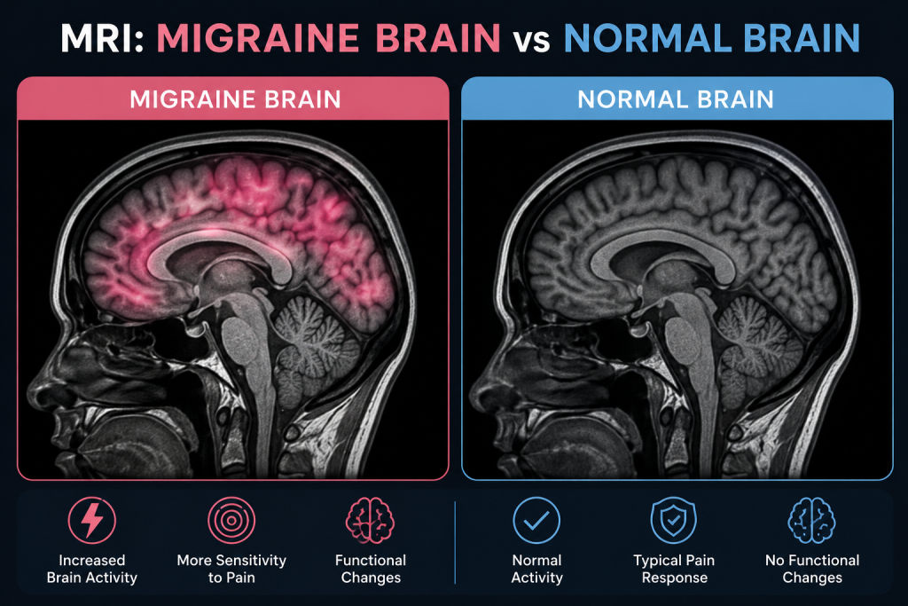
A brain MRI can feel like it should provide a simple visual answer to migraine: one scan should look like a migraine brain and another should look normal. In routine medical care, it usually does not work that way.

Migraine is primarily a disorder of brain function and sensory processing. A person can have frequent, disabling migraine attacks and still have a structurally normal MRI. When an MRI does show small bright areas in the white matter, those findings are often nonspecific and can occur in people with migraine, with aging, with vascular risk factors, or in people who have no headache disorder at all.

The practical difference between an MRI migraine brain vs normal brain is therefore not a single signature image. Doctors interpret the scan together with the headache pattern, neurologic examination, age, vascular risks, aura symptoms, and the size, shape, location, and behavior of any MRI findings.

KEY TAKEAWAYS

·        Most people with a typical migraine pattern and a normal neurologic examination do not need brain imaging solely to confirm migraine.

·        A routine structural MRI can be completely normal even when migraine attacks are severe or frequent.

·        Some people with migraine have small white matter hyperintensities, often described as tiny bright spots on T2 or FLAIR images.

·        A 2025 systematic review found that migraine-associated white matter findings are commonly small, supratentorial, and concentrated in deep frontal or parietal white matter, but their clinical significance remains uncertain.

·        White spots do not automatically mean multiple sclerosis, stroke, dementia, or a brain tumor. The MRI pattern and the clinical history determine what they mean.

·        Imaging becomes more important when a headache is sudden, new or changing, occurs with a neurologic deficit, starts after age 50, follows trauma, or appears with other red flags.

Think of the MRI as Three Possible Reports

A useful way to understand migraine imaging is to imagine three common report patterns rather than searching for one “migraine picture.”

MRI Pattern What the Radiologist May See How It Is Usually Interpreted
Report A: Structurally normal MRI No mass, acute stroke, bleeding, hydrocephalus, or significant white matter abnormality Very common in people with migraine; does not argue against a migraine diagnosis
Report B: A few small nonspecific white matter foci Tiny T2/FLAIR bright spots, often in deep or subcortical white matter Can be seen with migraine, aging, or vascular factors; interpreted by pattern and clinical context
Report C: Atypical or concerning pattern Mass effect, acute infarct, unusual enhancement, characteristic demyelinating pattern, or other focal abnormality May point to a diagnosis other than uncomplicated migraine and usually needs targeted follow-up

What Does a Normal Brain MRI Look Like in Someone With Migraine?

On a routine MRI, a “normal” brain means the radiologist does not see a structural abnormality that explains the symptoms. The brain tissue, ventricles, major fluid spaces, and visualized structures have an expected appearance for the person’s age, and there is no acute infarct, suspicious mass, major bleeding, or other significant finding.

That result is entirely compatible with migraine. MRI is excellent at showing anatomy, but a standard clinical scan is not designed to diagnose the moment-to-moment network activity that produces migraine pain, light sensitivity, nausea, aura, and sensory amplification.

Mayo Clinic explains this distinction clearly in its current migraine diagnosis guidance: migraine can involve abnormal brain function while the underlying structural MRI remains normal.

This is why a normal MRI should not be interpreted as “the headache is not real.” It means that the scan did not identify a structural cause for the symptoms.

What Are the White Spots Sometimes Seen on a Migraine MRI?

The most discussed structural MRI finding in migraine is the white matter hyperintensity, often abbreviated WMH. These are bright-appearing areas on certain MRI sequences, especially T2-weighted and FLAIR images.

A 2023 meta-analysis of 30 studies involving 3,502 people with migraine reported a pooled WMH prevalence of 44%. The pooled estimate was 45% in migraine with aura and 38% in migraine without aura. The studies were highly variable, so those percentages should not be used to predict whether one individual will have lesions.

More recent evidence remains cautious. A 2025 systematic review covering approximately 3,600 participants found that white matter findings were frequently reported in migraine, but the clinical meaning, progression, and relationship with migraine features remained uncertain. Across studies, the lesions were most often small, located above the tentorium, and concentrated in deep frontal or parietal white matter.

In other words, these spots can be associated with migraine, but they are not a migraine fingerprint. They are nonspecific imaging findings.

How Can Migraine White-Matter Spots Differ From Other MRI Lesions?

Radiologists do not judge a white spot by brightness alone. They look at its location, shape, number, size, whether it contacts the cortex or ventricles, whether it involves the corpus callosum, brainstem, or cerebellum, whether it restricts diffusion, and whether it enhances after contrast.

Possible Explanation Typical Imaging Clues Why Context Matters
Migraine-associated pattern Often small and punctate; commonly deep or subcortical frontal/parietal white matter Usually nonspecific; clinical context is essential
Age/small-vessel pattern Can involve deep and periventricular white matter; burden often increases with age and vascular risk May prompt review of blood pressure, diabetes, cholesterol, smoking, and vascular health
Multiple sclerosis pattern More likely to include lesions in characteristic periventricular, juxtacortical/cortical, infratentorial, corpus callosal, or spinal-cord locations Diagnosis requires the overall MRI pattern plus clinical and sometimes laboratory evidence
Acute ischemic stroke Often follows a vascular territory or shows restricted diffusion on appropriate sequences Requires urgent clinical correlation
Tumor or mass Usually masslike rather than a few scattered tiny WMHs; may cause swelling, mass effect, or enhancement Needs specialist evaluation based on the exact lesion type

A July 2026 Mayo Clinic guide to white spots on brain MRI emphasizes that white-matter spots may be linked with aging, small-vessel disease, migraine, stroke, multiple sclerosis, and other conditions. Location is useful, but it is only one part of the interpretation.

Does Migraine With Aura Look Different From Migraine Without Aura?

There is no routine MRI finding that reliably separates every person with aura from every person without aura. Population research suggests white matter lesions may be somewhat more common in migraine with aura, but an individual patient with aura can have a completely normal MRI, and a patient without aura can have small nonspecific spots.

Aura itself is a temporary neurologic phenomenon, most often visual but sometimes sensory or language-related. It is thought to involve cortical spreading depolarization, a wave of altered electrical and chemical activity across the cortex. A standard structural MRI is not a live map of that event.

Research MRI techniques such as functional MRI, perfusion imaging, diffusion methods, spectroscopy, or connectivity analysis can identify group-level differences in migraine studies. Those findings are scientifically important, but they are not currently a routine clinical test that lets a radiologist look at one scan and say, “this is definitely migraine.”

Can an MRI Tell Whether Migraine Has Damaged the Brain?

The presence of white matter spots often raises this fear, but the evidence does not support treating every migraine-associated WMH as brain damage from repeated attacks.

The American Migraine Foundation notes that these lesions are generally not linked to neurological impairment or cognitive decline in typical migraine patients. Research continues because migraine, especially migraine with aura, has associations with vascular risk in some populations, but an incidental small WMH is not the same as a clinical stroke.

The 2025 systematic review reached a similarly cautious conclusion: WMHs are common in migraine, yet their prognostic significance is still unclear, and their presence alone does not justify changing routine migraine management in an otherwise typical presentation.

Why Might a Doctor Order an MRI for a Headache?

The purpose of MRI is usually not to prove that migraine exists. It is to investigate features that make a secondary cause more plausible.

The current American College of Radiology Appropriateness Criteria for Headache state that imaging is usually not appropriate for a typical primary migraine or tension-type headache with a normal neurologic examination and no red flags. Imaging becomes appropriate in several higher-risk scenarios.

  • A first or worst headache, especially a sudden thunderclap headache that reaches maximum intensity rapidly.
  • A major change in headache frequency, severity, duration, or clinical pattern.
  • New weakness, numbness, trouble speaking, persistent visual loss, confusion, seizure, or another focal neurologic deficit.
  • New headache beginning after age 50.
  • Headache associated with fever, cancer, significant immunocompromise, or recent head trauma.
  • Features suggesting abnormal intracranial pressure, such as papilledema or certain positional symptoms.
  • New or unusual headache during pregnancy or the postpartum period.
  • Unusual, prolonged, or persistent aura, hemiplegic features, brainstem symptoms, or other atypical migraine presentations.

The exact test may be MRI, CT, vascular imaging, or a combination depending on the suspected problem. A sudden thunderclap headache, for example, is approached differently from a slowly changing chronic migraine pattern.

What Does “Nonspecific White Matter Change” Mean on an MRI Report?

The word nonspecific is important. It means the appearance does not point to one unique diagnosis by itself.

A report might say “a few scattered punctate T2/FLAIR hyperintensities in the subcortical white matter, nonspecific.” In a younger person with a long history of migraine and no neurologic deficit, a neurologist may consider the spots compatible with migraine. In an older adult with hypertension, diabetes, smoking history, or high cholesterol, similar spots may be interpreted in the context of small-vessel disease.

If the lesions have a distribution that is more typical of a demyelinating disease, the radiologist may specifically raise that possibility. The next step can include a neurologic examination, comparison with prior scans, spinal MRI, contrast-enhanced imaging, or laboratory testing. The report wording should be read as a description of probability and pattern, not as a diagnosis by itself.

MRI Migraine Brain vs MS Brain: Why the Difference Is Not Just “White Spots”

Migraine and multiple sclerosis can both be associated with white-matter lesions, which is why incidental spots sometimes cause understandable anxiety. But MS is diagnosed from a combination of clinical history and lesions that demonstrate characteristic distribution and, when applicable, dissemination in space and time.

MS lesions more often involve characteristic areas such as periventricular white matter, the cortex or juxtacortical region, the infratentorial brain, the corpus callosum, or spinal cord. Migraine-associated lesions described in research are more commonly small and deep or subcortical, often in the frontal regions.

There can be overlap, and no online checklist can replace a neuroradiologist or neurologist reviewing the actual images. A few nonspecific spots do not establish MS, and a normal brain MRI in a person with typical migraine is common.

What Questions Should You Ask After a Brain MRI?

If the report contains unfamiliar language, these questions are more useful than searching every word individually:

  • Was the MRI otherwise normal for my age?
  • Are the white-matter spots considered nonspecific, migraine-associated, vascular, or atypical?
  • Does the location or shape suggest multiple sclerosis or another demyelinating condition?
  • Is there evidence of an old or recent stroke?
  • Do I need contrast, vascular imaging, spinal MRI, or another test?
  • Should this MRI be compared with an older scan or repeated later?
  • Do the findings change my migraine treatment or my vascular-risk management?

A stable scan can also be useful. When prior imaging is available, radiologists can determine whether lesions are unchanged or whether there are genuinely new findings.

When Should a Headache Be Treated as an Emergency?

Seek emergency medical evaluation for a sudden severe headache that reaches peak intensity within seconds to minutes, especially if it is new. Also seek urgent care for headache with new weakness, facial droop, trouble speaking, fainting, severe confusion, seizure, new persistent vision loss, a stiff neck with fever, or symptoms after a significant head injury.

People who are pregnant or recently postpartum, are significantly immunocompromised, have active cancer, or develop a major new headache pattern may also need faster evaluation. The urgency comes from the symptoms and clinical setting, not from whether the person has a previous migraine diagnosis.

Frequently Asked Questions

Can a neurologist diagnose migraine from an MRI?

Usually no. Migraine is primarily diagnosed from symptoms, history, and a neurologic examination. MRI is mainly used when clinicians need to rule out another cause or investigate atypical features.

Does a normal MRI mean I do not have migraine?

No. A structurally normal MRI is common in migraine. Migraine can involve abnormal brain function without a visible structural abnormality on routine imaging.

Are white spots common with migraine?

They can be. Studies report variable rates, and a 2023 meta-analysis estimated a pooled prevalence of about 44% among people with migraine. The spots are nonspecific and are not present in everyone with migraine.

Do migraine white spots turn into multiple sclerosis?

Migraine-associated white matter spots do not simply turn into MS. MS is a separate disease diagnosed from its clinical presentation and characteristic imaging and laboratory evidence.

Can MRI distinguish migraine aura from a stroke?

MRI can help evaluate suspected stroke, but the answer depends on timing, MRI sequences, symptoms, and other tests. A first episode of new weakness, speech trouble, or persistent visual loss should be treated urgently rather than assumed to be aura.

Do I need another MRI every year if I have migraine?

Usually not when migraine symptoms are stable, the neurologic examination is normal, and a prior MRI was reassuring. Repeat imaging is driven by new clinical features, concerning findings, or a specific monitoring plan.

Can a brain tumor be mistaken for migraine?

Some tumors can cause headache, but a tumor usually produces a structural abnormality on imaging rather than the tiny nonspecific white-matter spots commonly discussed in migraine. New neurologic deficits or a major change in headache pattern warrant medical evaluation.

The Bottom Line

When comparing an MRI migraine brain vs normal brain, the most important fact is that the two can look exactly the same on a routine structural scan. Migraine is not diagnosed by finding one characteristic lesion.

Some people with migraine have small white matter hyperintensities, often in deep or subcortical frontal and parietal regions. These findings are usually nonspecific, and current research still does not establish that they represent clinically meaningful brain damage in a typical migraine patient.

What matters most is the pattern. A radiologist considers lesion size, location, shape, enhancement, diffusion, age, vascular risk factors, symptoms, and prior scans. If the headache pattern is typical and the neurologic examination is normal, major imaging abnormalities are uncommon. If the symptoms are new, sudden, changing, or neurologically unusual, imaging can become an important part of ruling out a secondary cause.

Medical note: This article provides general educational information and is not a diagnosis. Brain MRI findings must be interpreted by a qualified clinician or radiologist in the context of symptoms, examination, age, health history, and prior imaging. Evidence and imaging guidance were checked against current U.S. sources available in August 2026.

A practical diagnostic pathway for infections, dental causes, salivary gland problems, persistent neck masses, testing, and treatment in 2026.

Finding a lump beneath the jaw can be unsettling. A swollen submandibular lymph node often means the immune system is reacting to a nearby problem, such as a sore throat, cold, skin irritation, or dental infection. The location matters because these nodes drain areas of the mouth, lower face, tongue, gums, and nearby tissues.

Still, not every lump in this area is a lymph node. The submandibular salivary gland sits nearby and can enlarge when a duct is blocked by a stone or when the gland becomes infected. The most useful first step is therefore not guessing the diagnosis, but following the clues: where the lump sits, whether it hurts, how quickly it appeared, what other symptoms are present, and whether it is shrinking or persisting.

KEY TAKEAWAYS

·        Submandibular nodes commonly enlarge because of infections in the teeth, gums, throat, mouth, or nearby skin.

·        A tender, movable node that appears with an obvious infection is usually less concerning than a hard, fixed, painless, or steadily enlarging mass.

·        Adults should not ignore a neck mass that persists without a clear infectious cause, especially when it lasts several weeks or comes with swallowing, voice, mouth, or weight-loss symptoms.

·        Children often develop reactive neck nodes during routine infections, but nodes larger than about 2 centimeters, matted nodes, or persistent unexplained swelling deserve evaluation.

·        Treatment targets the underlying cause. Antibiotics, steroids, or repeated squeezing should not be used as do-it-yourself treatments for an unexplained lump.

First question: Is the lump actually a lymph node?

The term submandibular means “under the lower jaw.” Several structures share this small space, so a lump can come from different tissues. A clinician distinguishes them by location, movement, consistency, and what makes the swelling change.

What it may be Clues that point in that direction
Reactive lymph node Usually a small oval or bean-shaped lump. It may be tender and mobile, especially during a cold, sore throat, dental problem, or skin infection.
Submandibular salivary gland or stone Often feels broader or deeper than a node. Pain or swelling may increase around meals, when saliva production rises.
Dental abscess or jaw infection May occur with toothache, gum swelling, bad taste, facial swelling, fever, or pain when chewing.
Skin cyst Often sits closer to the skin surface and may have a visible pore, redness, or a history of recurring inflammation.
Other neck mass A persistent, firm, fixed, or enlarging lump may require imaging and tissue sampling to identify its origin.

Why do submandibular lymph nodes swell?

Lymph nodes act as immune checkpoints. They filter lymphatic fluid and help immune cells recognize germs or abnormal cells. When tissue in the drainage area becomes inflamed, the nearby nodes may enlarge as immune activity increases. The swelling itself is a sign, not a disease.

The dental and mouth pathway

Teeth and gums are especially important when a node under the jaw becomes tender on one side. Tooth decay, a dental abscess, infected gums, recent dental procedures, mouth ulcers, and infections around a partially erupted wisdom tooth can all trigger nearby nodes. A dental source becomes more likely when there is tooth sensitivity, pain with biting, gum swelling, facial pressure, pus, a foul taste, or reduced ability to open the mouth.

The throat, nose, and skin pathway

Viral upper respiratory infections, strep throat, tonsillitis, influenza, and other infections can cause one or several cervical nodes to react. Cuts, acne, shaving irritation, or infected skin on the lower face may do the same. Direct infection of a lymph node, called lymphadenitis, is more likely when the node is very painful and the overlying skin becomes red, hot, or swollen. Fever may be present, and an abscess can occasionally form.

The bodywide pathway

When lymph nodes are enlarged in several regions, clinicians consider systemic infections and inflammatory conditions. Examples include infectious mononucleosis, cytomegalovirus, HIV, tuberculosis, cat-scratch disease, toxoplasmosis, autoimmune illnesses, and some medication reactions. Travel, animal exposure, recent vaccines, new prescriptions, immune status, and sexual history can all affect the diagnostic plan.

The less common but important pathway

Cancer is not the most common explanation for a submandibular lump, but persistent neck masses require appropriate attention. Lymphoma can begin in lymphatic tissue, while cancers of the mouth, tongue, throat, salivary gland, or other nearby structures may spread to cervical nodes. Risk rises with age and with exposures such as tobacco and heavy alcohol use, but serious causes can occur in people without those risk factors.

A 2025 review in American Family Physician recommends evaluating the location, size, texture, mobility, tenderness, duration, systemic symptoms, exposures, medications, and whether the swelling is localized or generalized. It also notes that persistent lymphadenopathy beyond four weeks or swelling accompanied by systemic symptoms may require laboratory testing and imaging.

What do the feel and timing of the node suggest?

Finding What it may mean
Tender and appeared quickly Often fits an infection or inflammation, particularly when there is a sore throat, tooth problem, fever, or skin irritation.
Soft or rubbery and movable Can occur with reactive nodes. “Movable” is reassuring only when the overall history also looks benign.
Hard, fixed, or matted Needs medical evaluation because these features can occur with malignancy or granulomatous disease.
Painless and steadily enlarging More concerning than a node that becomes tender during a clear infection and then shrinks.
Fluctuant, red, and hot May indicate suppurative lymphadenitis or an abscess that needs prompt treatment.
Present in several body regions Suggests a systemic process rather than a single local dental or throat problem.

Important: Texture and tenderness cannot diagnose the cause on their own. A clinician considers the complete pattern and may use imaging or biopsy when needed.

When should you make an appointment?

Arrange a medical or dental evaluation when the lump has no obvious explanation, is getting larger, or does not begin to settle after the suspected infection improves. The threshold for evaluation should be lower in adults with a new persistent neck mass.

  • The lump remains present for several weeks or keeps returning.
  • It is hard, fixed, matted to nearby tissue, or clearly enlarging.
  • It is larger than expected, especially around 2 centimeters or more in a child.
  • There are drenching night sweats, unexplained fever, persistent fatigue, or unintentional weight loss.
  • You have a mouth sore, tongue lesion, hoarseness, ear pain without an ear infection, difficulty swallowing, or a change in voice that does not resolve.
  • You have ongoing tooth pain, gum swelling, facial swelling, or signs of a dental abscess.
  • Nodes are enlarged in the armpits, groin, or several areas in addition to the neck.

The American Academy of Otolaryngology-Head and Neck Surgery guideline emphasizes prompt, accurate evaluation of adult neck masses when malignancy is possible, rather than allowing repeated empiric treatment to delay diagnosis.

When is swelling under the jaw urgent?

Seek urgent care the same day for rapidly worsening pain, high fever, spreading redness, pus, significant facial swelling, dehydration, or an inability to open the mouth normally. Go to an emergency department for trouble breathing, drooling with inability to swallow, severe neck stiffness, swelling that pushes the tongue upward, confusion, fainting, or other signs of a deep neck infection or airway problem.

What happens during the medical evaluation?

The workup usually follows a sequence rather than jumping immediately to biopsy. The purpose is to identify an obvious local cause while recognizing features that require faster investigation.

  1. History: The clinician asks when the lump appeared, whether it is changing, recent illnesses, dental symptoms, pain with meals, travel, animal contact, medication changes, vaccines, immune conditions, tobacco or alcohol exposure, and symptoms such as fever, night sweats, or weight loss.
  2. Focused examination: The mouth, teeth, gums, tongue, tonsils, skin, salivary glands, ears, and neck are examined. The clinician checks node size, tenderness, texture, mobility, and whether other lymph node regions are involved.
  3. Targeted tests: A complete blood count, inflammatory markers, throat testing, viral testing, tuberculosis testing, or other labs may be chosen based on the history. Not everyone needs the same panel.
  4. Imaging: Ultrasound is commonly the first imaging test for peripheral lymphadenopathy in children age 14 and younger. Adults with persistent or higher-risk neck lymphadenopathy may need contrast-enhanced CT or MRI.
  5. Tissue sampling: Fine-needle aspiration, core needle biopsy, or excisional biopsy may be used when imaging is suspicious, the cause remains unclear, or the node does not resolve as expected. Core biopsy can provide more tissue architecture than a simple aspiration.

The Merck Manual overview of lymphadenopathy notes that many people with newly identified localized nodes and no red flags can be observed for up to about four weeks, while persistent unexplained nodes, generalized swelling, or concerning features call for further testing. Observation is a clinical decision, not a reason to ignore changes.

How is a swollen submandibular lymph node treated?

There is no medication that treats “a swollen node” as a stand-alone diagnosis. Treatment is directed at the cause.

When the cause is viral

Supportive care may be enough for an uncomplicated viral illness. Rest, appropriate fluids, and over-the-counter pain or fever medication may help when safe for the individual. The node can remain noticeable for a while after the infection improves because immune tissue may shrink gradually.

When the cause is bacterial

Antibiotics are used when a clinician identifies or strongly suspects a bacterial infection, such as bacterial lymphadenitis, strep throat, or a dental infection. An abscess may require drainage. Antibiotics do not treat viral illness, lymphoma, salivary stones, or other nonbacterial causes, so taking leftover medication can delay the correct diagnosis.

When the cause is dental

Dental treatment may involve drainage, root canal therapy, extraction, periodontal care, or antibiotics when the infection has spread or systemic symptoms are present. Pain relief alone does not remove the source of an abscess. Facial swelling, fever, or difficulty swallowing requires prompt attention.

When the cause is a salivary stone

Management may include hydration, warm compresses, gentle gland massage, and measures that stimulate saliva, but only after a clinician confirms the likely problem and checks for infection. Recurrent obstruction, severe pain, or infection may require an ear, nose, and throat specialist or oral surgeon.

When the cause is inflammatory or malignant

Autoimmune disease, granulomatous disease, lymphoma, leukemia, and metastatic cancer require condition-specific treatment. Corticosteroids should generally not be started for unexplained lymphadenopathy before diagnosis because they can temporarily shrink lymphoma-related nodes and interfere with tissue interpretation.

What should you avoid doing at home?

  • Do not repeatedly squeeze, dig at, or aggressively massage an unidentified lump.
  • Do not start leftover antibiotics or someone else’s prescription.
  • Do not take corticosteroids simply to make the node shrink before the cause is known.
  • Do not assume that a painless lump is harmless because it does not hurt.
  • Do not rely on size alone. Trend, texture, duration, symptoms, age, and location all matter.

How long can a reactive node stay enlarged?

A node caused by a routine infection may start to soften and shrink as the illness resolves, but it can remain palpable after other symptoms disappear. What matters is the direction of change. A node that is steadily getting smaller is different from one that is unchanged, enlarging, recurring, or becoming firmer. Persistent swelling beyond roughly four weeks, especially without a clear cause or with systemic symptoms, generally warrants reassessment.

Does age change the level of concern?

Yes. Children frequently develop reactive cervical nodes because their immune systems encounter many respiratory and throat infections. Ultrasound is often favored first when imaging is needed. Even so, a very large node, hard or matted nodes, prolonged unexplained swelling, supraclavicular nodes, or systemic symptoms should not be dismissed.

In adults, a persistent neck mass deserves more deliberate evaluation because the probability of malignancy increases with age. A new lump that lasts, enlarges, or appears without an obvious infection should be assessed rather than repeatedly treated as “swollen glands.”

Questions worth bringing to the appointment

  • Does this feel like a lymph node, salivary gland, cyst, or another type of mass?
  • Is there evidence of a dental, throat, skin, or salivary infection?
  • What change should I expect, and by what date should it be smaller?
  • Which symptoms would mean I need urgent care?
  • Do I need ultrasound, CT, MRI, blood tests, dental imaging, or a biopsy?
  • Should I see a dentist, primary care clinician, pediatrician, ENT specialist, hematologist, or oral surgeon?

The practical bottom line

Most swelling in a submandibular lymph node is reactive and linked to a nearby infection, particularly in the mouth, teeth, throat, or skin. Tenderness, mobility, a clear infectious trigger, and gradual improvement are generally reassuring features. They do not replace evaluation when the pattern is unusual.

A hard, fixed, painless, steadily enlarging, or persistent lump should be examined, as should swelling associated with night sweats, unexplained weight loss, mouth or throat symptoms, widespread nodes, or significant dental or facial infection. The right next step may be simple observation, dental care, laboratory testing, imaging, or tissue sampling. The goal is not to fear every lump, but to avoid overlooking one that is behaving differently from a routine reactive node.

Medical note: This article is for general educational purposes and does not diagnose a neck lump or replace care from a licensed healthcare professional. Seek urgent help for breathing or swallowing difficulty, rapidly spreading swelling, or severe illness.

An evidence-based look at the thyroid condition she discussed publicly, the symptoms that can be missed, and the limits of diagnosing anyone through headlines or appearance.

The phrase Wendy Williams Graves disease became widely searched after the television host told viewers in 2018 that she was living with Graves’ disease and hyperthyroidism. Her disclosure brought unusual public attention to an autoimmune thyroid disorder that can affect heart rate, sleep, mood, weight, muscles, eyes, and day-to-day stamina.

Her story is useful as an awareness starting point, but it is not a medical chart. Public appearances cannot show whether Graves’ disease is active, in remission, controlled by medication, or connected to any later health development. The safest way to understand the condition is to separate verified public statements from general medical facts.

Key Takeaways

Wendy Williams publicly disclosed Graves’ disease and hyperthyroidism in 2018. Graves’ disease is an autoimmune cause of an overactive thyroid, not simply a “fast metabolism.” Symptoms can affect many body systems and may be mistaken for stress, menopause, anxiety, or overwork. Treatment may involve antithyroid medication, radioactive iodine, surgery, and symptom control. Eye disease can follow a different course from thyroid hormone levels. Her current thyroid status is private and cannot be determined from media coverage.

The Public Moment That Changed the Conversation

On February 21, 2018, Williams announced on her daytime program that she had Graves’ disease and hyperthyroidism and would take a medically recommended break. Contemporary reporting noted that her team said she had been dealing with Graves’ disease for years. ABC News documented that announcement, including her description of thyroid-related eye changes and the need to get her medication and hormone levels back in balance.

That disclosure mattered because Graves’ disease often looks nonspecific from the outside. A person may be exhausted but unable to sleep, lose weight while eating normally, feel overheated, experience a racing heartbeat, or become unusually irritable. Those changes can be explained away as work stress or aging until blood tests reveal an overactive thyroid.

What Is Publicly Known, and What Is Not?

Supported by public reporting Not possible to conclude from public reporting
Williams said she had Graves’ disease and hyperthyroidism. Whether her Graves’ disease is currently active, controlled, or in remission.
She publicly connected eye changes and a need for medical leave with her thyroid condition. Whether any later symptom, diagnosis, or career event was caused by Graves’ disease.
She has also separately discussed lymphedema. That lymphedema and Graves’ disease are the same condition or have the same cause.

Graves’ Disease Is an Immune Signal Problem

The thyroid is a small gland at the front of the neck, but its hormones influence nearly every organ. In Graves’ disease, the immune system produces antibodies that act like a stuck accelerator. These antibodies stimulate the thyroid to release more hormone than the body needs. The result is hyperthyroidism, a state in which many body processes run too fast.

The National Institute of Diabetes and Digestive and Kidney Diseases estimates that Graves’ disease affects nearly 1 in 100 Americans and causes about 4 out of 5 cases of hyperthyroidism in the United States. It is more common in women and in people older than 30, although it can occur at other ages.

The “Signal Map”: How an Overactive Thyroid Can Show Up

Body system Possible signs Why it may be overlooked
Heart and circulation Fast pulse, pounding heartbeat, irregular rhythm, higher blood pressure May be blamed on caffeine, panic, or poor conditioning
Nervous system Tremor, nervousness, restlessness, irritability, trouble sleeping May resemble anxiety or chronic stress
Metabolism Weight loss, heat intolerance, sweating, increased appetite Weight change may be welcomed instead of investigated
Muscles and bones Weakness, fatigue, reduced exercise tolerance, bone loss over time Fatigue may be blamed on age or a busy schedule
Digestive and reproductive systems Frequent bowel movements, menstrual changes, fertility concerns Symptoms may be treated separately
Eyes Dryness, grittiness, swelling, light sensitivity, bulging, double vision Eye symptoms may be mistaken for allergies or lack of sleep

Why Did Her Eyes Become Part of the Discussion?

Graves’ disease can affect tissue around the eyes, producing thyroid eye disease, also called Graves’ ophthalmopathy. This is not simply a cosmetic change. Inflammation can cause dry or gritty eyes, eyelid swelling, pressure, light sensitivity, bulging, pain with eye movement, or double vision. Rarely, severe swelling can threaten vision.

The eye condition and the thyroid condition are related, but they do not always move together. Eye symptoms can begin before hyperthyroidism is diagnosed, appear during treatment, or continue after thyroid hormone levels normalize. Smoking and nicotine exposure are especially important because they increase the risk and severity of thyroid eye disease.

Urgent eye warning

New loss of color vision, reduced vision, severe eye pain, inability to close the eyelids, or rapidly worsening double vision deserves prompt medical assessment. These are not symptoms to monitor through photographs or social media commentary.

How Doctors Confirm Graves’ Disease

A clinician does not diagnose Graves’ disease from weight, behavior, eye appearance, or a single symptom. The evaluation usually combines a medical history, a neck and eye examination, pulse and blood pressure checks, and laboratory testing.

  • TSH is usually low when the thyroid is producing too much hormone.
  • Free T4 and sometimes T3 help show the degree of thyroid hormone excess.
  • Thyroid-stimulating immunoglobulin or TSH receptor antibody testing can support a Graves’ disease diagnosis.
  • A radioactive iodine uptake test or thyroid scan may show diffuse overactivity throughout the gland.
  • Doppler ultrasound may be used when radioactive iodine testing is not appropriate, including in pregnancy or breastfeeding.

Testing also helps separate Graves’ disease from thyroiditis, toxic thyroid nodules, medication effects, and other causes of hyperthyroidism. That distinction matters because the treatments are not interchangeable.

Treatment Has Three Different Jobs

1. Calm the immediate symptoms

Beta-blockers may be prescribed to reduce a racing heart, tremor, and nervousness while thyroid-specific treatment begins to work. They do not stop the thyroid from producing excess hormone, but they can make a patient feel better relatively quickly.

2. Reduce or remove excess thyroid hormone production

The main treatment routes are antithyroid medication, radioactive iodine, and thyroid surgery. The American Thyroid Association’s Graves’ disease guidance explains that methimazole is commonly used, while propylthiouracil is reserved for certain situations. Radioactive iodine gradually destroys overactive thyroid cells. Thyroidectomy removes most or all of the gland and is often considered when a large goiter, suspicious nodule, medication problem, pregnancy-related issue, or severe eye disease changes the risk-benefit calculation.

Radioactive iodine and surgery frequently lead to hypothyroidism. That outcome is expected and treated with daily thyroid hormone replacement. It does not mean treatment failed.

3. Protect the organs affected by the disease

Treatment may also address thyroid eye disease, bone health, heart rhythm, pregnancy planning, or severe muscle weakness. Eye care can range from lubricating drops and smoking cessation to immune-directed medication, radiation in selected cases, or surgery. Management should be coordinated with an endocrinologist and, when needed, an ophthalmologist who has experience with thyroid eye disease.

Why Medical Leave Can Help, but Rest Is Not the Cure

Williams’ temporary leave highlighted an important reality: uncontrolled hyperthyroidism can make normal work demands difficult. Rest can reduce strain while medication is adjusted, sleep improves, and heart rate comes under control. Still, Graves’ disease is not cured by taking time off. The immune-driven thyroid overactivity requires medical monitoring and, in many cases, long-term treatment.

Patients also need repeat blood tests because symptoms may improve before hormone levels fully normalize, or laboratory values may change before the person notices a difference. Medication doses that are too high can shift someone into hypothyroidism, which has its own symptoms and risks.

Graves’ Disease and Lymphedema Should Not Be Blended Together

Williams has publicly discussed both Graves’ disease and lymphedema. They are separate medical conditions. Graves’ disease is an autoimmune thyroid disorder. Lymphedema is swelling caused by impaired movement of lymph fluid. A rare Graves-related skin condition called pretibial myxedema can cause thickened, reddish skin on the shins or feet, but it is not a general explanation for every form of leg or ankle swelling.

This distinction matters for readers searching celebrity health stories. Similar-looking symptoms do not prove a shared cause, and one diagnosis should not be used to reinterpret every later symptom.

Can Graves’ Disease Go Away?

Some people enter remission after a course of antithyroid medication, meaning thyroid function remains normal after the drug is stopped. Others relapse months or years later. Factors such as antibody levels, thyroid size, smoking, disease severity, and treatment history can influence the likelihood of remission, but no public image or interview can reveal an individual patient’s status.

After radioactive iodine or complete thyroid removal, the thyroid can no longer overproduce hormone in the same way, but lifelong thyroid hormone replacement is usually needed. Thyroid eye disease may still require separate follow-up because it can remain active even when thyroid blood tests are controlled.

Five Lessons From Wendy Williams’ Disclosure

A busy life can hide a medical problem. Insomnia, irritability, sweating, fatigue, and weight change may be rationalized until several symptoms form a pattern.

Eye changes deserve medical attention. Dryness, swelling, bulging, or double vision can be part of thyroid eye disease and should not be dismissed as appearance alone.

“Hyperthyroidism” and “Graves’ disease” are related, but not identical terms. Hyperthyroidism describes excess thyroid hormone. Graves’ disease is one autoimmune cause of it.

Visible improvement does not prove remission. Only clinical follow-up and laboratory testing can show whether thyroid activity is controlled.

Celebrity disclosure can raise awareness, but it cannot replace individual care. Readers should use the story as a prompt to recognize symptoms, not as a template for self-diagnosis.

When Should Someone Ask for a Thyroid Evaluation?

A primary care clinician or endocrinologist may consider thyroid testing when several symptoms occur together, especially a fast or irregular heartbeat, unexplained weight loss, heat intolerance, tremor, neck swelling, persistent insomnia, unusual weakness, or new eye symptoms. People with a family history of autoimmune thyroid disease or another autoimmune condition may have a higher index of suspicion.

Emergency symptoms

Call 911 for chest pain, fainting, severe shortness of breath, a very rapid or irregular heartbeat with weakness, new confusion, high fever, or severe agitation. Rarely, uncontrolled hyperthyroidism can escalate into thyroid storm, a life-threatening emergency.

Questions Readers Commonly Ask

Did Graves’ disease cause all of Wendy Williams’ later health problems?

That cannot be concluded from public information. She has discussed more than one health condition, and only her treating clinicians can determine whether any conditions are related.

Is Graves’ disease caused by stress?

Stress may affect symptoms or act as a trigger in a susceptible person, but Graves’ disease is an autoimmune disorder influenced by multiple genetic and environmental factors. Stress alone is not a complete explanation.

Can Graves’ disease change personality or mood?

Excess thyroid hormone can contribute to irritability, anxiety, restlessness, poor sleep, and difficulty concentrating. These symptoms should be evaluated medically rather than assumed to be a personality change.

Does everyone with Graves’ disease develop bulging eyes?

No. Many people never develop clinically significant thyroid eye disease. Others have mild dryness or swelling, while a smaller group develops more visible or serious symptoms.

Should people with Graves’ disease avoid iodine?

Large iodine exposures, including some supplements and seaweed products, can worsen hyperthyroidism in susceptible people. Patients should discuss supplements, multivitamins, contrast imaging, and dietary changes with their clinician rather than adopting a restrictive diet on their own.

The Bottom Line

Wendy Williams helped bring Graves’ disease into mainstream conversation by speaking publicly about her diagnosis, eye symptoms, and need for medical leave. The larger lesson is not to diagnose her current health from a distance. It is to recognize that an autoimmune thyroid disorder can create widespread symptoms, that eye disease may require separate care, and that effective treatment depends on laboratory monitoring and an individualized plan.

Anyone who recognizes a cluster of symptoms should seek a proper thyroid evaluation rather than relying on a celebrity comparison. Graves’ disease is treatable, but untreated hyperthyroidism can affect the heart, bones, muscles, fertility, pregnancy, and vision.

Authoritative external resources

ABC News: Wendy Williams’ 2018 public disclosure. National Institute of Diabetes and Digestive and Kidney Diseases: Graves’ disease symptoms, diagnosis, and treatment. American Thyroid Association: patient guidance on Graves’ disease and treatment options.

Medical disclaimer: This article is for general educational purposes. It does not diagnose Wendy Williams or any reader, and it does not replace care from a qualified healthcare professional.

Brand prices, generic options, insurance costs, and practical ways to pay less in the United States

August 2026 price snapshot
A brand-name EpiPen 2-Pak can still carry a retail price near $650 to $750 before insurance or discounts. Generic and authorized-generic epinephrine auto-injectors are often available for roughly $175 to $300 per two-pack when a current pharmacy coupon is used. Your actual cost may be much lower with insurance or a manufacturer program.

The question “how much does an EpiPen cost?” sounds as though it should have one simple answer. In practice, the number at the pharmacy can change by hundreds of dollars depending on the product written on the prescription, the pharmacy, the insurance formulary, the deductible, and whether a savings card or cash coupon is used.

EpiPen is a brand name for an epinephrine auto-injector. Many people use the name for all emergency epinephrine devices, but the brand, authorized generic, FDA-approved generics, and competing devices do not necessarily have the same price or the same operating steps. That distinction matters when comparing costs.

Key Takeaways

  • A brand-name EpiPen 2-Pak may retail for about $650 to $750 without insurance, although the price at a specific pharmacy can be higher or lower.
  • Generic or authorized-generic epinephrine auto-injectors commonly fall near $175 to $300 for a two-pack with a current discount coupon.
  • Commercial insurance can reduce the cost to a low copay, but a deductible, coinsurance, or nonpreferred formulary tier may leave a much larger bill.
  • Eligible commercially insured patients may qualify for an EpiPen savings card worth up to $300 per carton, subject to current program terms.
  • Do not choose a device based only on price. Make sure the prescription, dose, training, and device design are appropriate for the person who will use it.

The EpiPen Price Ladder: What Might You Actually Pay?

The most useful way to understand the cost is to separate the sticker price from the final out-of-pocket price. The ranges below are national estimates and discount snapshots, not guaranteed pharmacy quotes.

Payment route Typical 2026 range What changes the price Best first step
Brand EpiPen, no insurance About $650 to $750 per 2-Pak before discounts Pharmacy, ZIP code, supply, and coupon availability Compare the brand price with the authorized generic before paying
Generic or authorized generic, cash coupon Often about $175 to $300 per two-pack Manufacturer, pharmacy, strength, and coupon code Ask the prescriber to permit an appropriate generic and compare pharmacies
Commercial insurance $0 or a low copay for some plans, but potentially hundreds with a deductible Formulary tier, deductible, coinsurance, prior authorization Check the exact National Drug Code or product name with the insurer and pharmacist
Medicare or Medicaid Plan and state specific Covered product, tier, deductible, and program rules Use the plan formulary and ask about the preferred epinephrine product
Manufacturer assistance Potentially much lower, including free medicine for some eligible patients Insurance type, income, residency, and program limits Review official eligibility rules before the prescription is filled

Why Can the Same EpiPen Prescription Produce Two Very Different Bills?

The price shown in an online search is not always the amount that an insured patient pays. Pharmacies can process the prescription through an insurance plan, a manufacturer savings card, or a prescription discount program. Those routes use different negotiated prices and usually cannot be stacked together.

1. Brand Versus Generic Wording

A prescription written specifically for brand EpiPen may be processed differently from one that allows generic substitution. Viatris sells an authorized generic that uses the same drug and device design under an epinephrine label rather than the EpiPen name. Other FDA-approved generic epinephrine auto-injectors may also be available, but devices can differ in appearance and instructions.

2. The Insurance Formulary

An insurance company may prefer one epinephrine product and place another on a higher tier. A preferred generic might have a small copay, while brand EpiPen may require prior authorization or full payment until a deductible is met. The cheapest retail product is not always the cheapest insured product, so both prices should be checked.

3. Pharmacy and ZIP Code

Cash and coupon prices can vary among pharmacies in the same city. Discount prices also change over time. A price found several months ago may not be honored at the next refill, which is why it is important to run a new comparison for the correct strength and quantity before every purchase.

4. Dose and Package Size

EpiPen 0.3 mg is generally used for people who weigh 66 pounds or more, while EpiPen Jr 0.15 mg is generally used for people who weigh 33 to 66 pounds. The prescription is usually filled as a carton containing two single-use devices. Comparing a one-device figure with a two-pack price can make one option appear cheaper than it actually is.

How Much Is EpiPen Without Insurance?

A patient paying the full brand-name retail price may see a bill around $650 to $750 for an EpiPen 2-Pak. This is the high end of the market and should not be treated as the only available price. Before paying, ask the pharmacist to quote the authorized generic and any FDA-approved generic listed on the prescription.

Current discount listings often place generic epinephrine near $175 to $300 per two-pack, with some widely available coupon prices around the mid-$170s. These numbers can move from week to week. The most reliable approach is to compare the exact prescription at nearby pharmacies on the day it will be filled.

For a live local comparison, check the current GoodRx epinephrine auto-injector price page and confirm the displayed product, strength, quantity, manufacturer, and pharmacy before presenting the coupon.

How Much Does EpiPen Cost With Insurance?

There is no universal insured price. A person whose plan treats generic epinephrine as a preferred drug may pay nothing or a modest copay. Another person with a high-deductible plan may pay most of the negotiated price early in the year. Coinsurance can also create a percentage-based charge rather than a fixed copay.

Before the prescription is sent, call the insurer or use its online drug-price tool. Ask which epinephrine auto-injector is preferred, whether prior authorization is required, what the two-pack will cost at a retail pharmacy, and whether a mail-order option is allowed. Give the prescriber the preferred product name so the pharmacy does not have to restart the process after a rejection.

Can an EpiPen Savings Card Lower the Cost?

Viatris currently offers savings programs for brand EpiPen and its authorized generic. Eligible commercially insured patients may save up to $300 on each EpiPen or EpiPen Jr 2-Pak carton, with use limits that apply during the calendar year. The authorized-generic card has a smaller maximum benefit. Program terms can change, and government-funded insurance such as Medicare, Medicaid, and TRICARE is generally excluded from manufacturer copay cards.

Review eligibility and current limits on the official EpiPen access and savings programs page before relying on a coupon amount shown on an older article or saved image.

People without prescription coverage or with financial hardship may also qualify for the Viatris Patient Assistance Program. Approval is not automatic. The program may require proof of U.S. residence, income information, a valid prescription, and participation by the healthcare professional.

Are Cheaper EpiPen Alternatives the Same Thing?

Every emergency epinephrine product delivers epinephrine, but the devices are not interchangeable in the everyday sense. The cap, needle end, activation method, hold time, voice prompts, dose options, and storage instructions can differ. A lower price is helpful only when the patient and caregivers know exactly how to use that device.

Option Potential cost advantage Important distinction
Authorized generic EpiPen Often much less than the brand cash price Same drug and device design under a generic label
Other generic epinephrine auto-injectors Coupon prices may compete with or beat the authorized generic Instructions may differ, so retraining is essential
AUVI-Q Most commercially insured patients may pay $35 or less; other program limits apply Compact device with voice instructions and different dose options
neffy nasal spray Eligible commercially insured patients may pay as little as $0 under current terms Prescription nasal epinephrine, not an auto-injector; coverage and clinical suitability vary

A prescriber may need to issue a new prescription when changing to another device. Ask the pharmacist for a trainer or demonstration and make sure family members, school staff, and caregivers are familiar with the product that was actually dispensed.

A Five-Minute Cost Check Before You Fill the Prescription

  1. Read the prescription name and dose. Confirm whether it says brand EpiPen, epinephrine auto-injector, EpiPen Jr, or another device.
  2. Check the insurance formulary. Find the preferred product and estimated cost for a two-device carton.
  3. Compare the insurance price with a cash coupon. A coupon may be cheaper, but purchases outside insurance may not count toward the deductible.
  4. Check the manufacturer program. Confirm eligibility, annual limits, expiration, and whether commercial insurance is required.
  5. Call the pharmacy. Ask whether the exact product is in stock and whether the quoted price matches the National Drug Code they will dispense.
  6. Ask for training. Switching devices to save money should always include updated instructions and practice with a trainer.

What Should Families Budget for Multiple Locations?

The cost can multiply when a child or adult needs epinephrine at home, school, work, daycare, or during travel. However, separating a two-pack may conflict with the instruction to keep two doses available because a second dose can be needed before emergency help arrives. Discuss the number of cartons needed with the prescriber rather than deciding solely on price.

Schools may stock undesignated epinephrine under state laws and local policies, but families should not assume the school supply replaces the student’s personal prescription. Ask the school nurse what is stocked, where it is kept, who may administer it, and whether a current individual action plan is required.

Do EpiPens Expire, and Does Replacement Affect the Annual Cost?

Epinephrine auto-injectors have expiration dates and must be replaced. Keep the devices at room temperature, protect them from light and extreme heat or cold, and inspect the solution when the device has a viewing window. A device that is expired, damaged, discolored, or contains particles should be replaced.

Because refills are recurring, the annual cost may include more than one carton. Set reminders several weeks before expiration so there is time to request a new prescription, compare prices, obtain prior authorization, and locate stock. Do not wait until the last device expires to begin the process.

Cost Should Never Delay Emergency Treatment

Epinephrine is the first-line emergency treatment for anaphylaxis. Use the prescribed device promptly when a serious allergic reaction is suspected, then follow the emergency plan and obtain medical help. Do not save a device for later because it was expensive.

The MedlinePlus epinephrine injection guide advises keeping two automatic injection devices available because one dose may not be enough. It also explains that epinephrine does not replace emergency medical care.

Frequently Asked Cost Questions

Is a Generic EpiPen as Effective as the Brand?

FDA-approved generic epinephrine auto-injectors must meet applicable standards for safety, effectiveness, quality, and device performance. An authorized generic EpiPen uses the same drug and device design as the brand product. Still, the label and appearance can differ, and another manufacturer’s device may use different operating steps.

Can I Use Insurance and a GoodRx Coupon Together?

Usually, no. The pharmacy generally processes the prescription through either insurance or the discount coupon. Compare both prices before choosing. A cash coupon purchase may not count toward an insurance deductible or out-of-pocket maximum.

Why Did the Pharmacy Price Change Since My Last Refill?

Discount contracts, pharmacy pricing, product availability, insurance formularies, and deductibles can change. Always generate a new coupon and reconfirm the exact product before each refill instead of relying on a screenshot from the previous purchase.

Can I Buy One EpiPen Instead of a Two-Pack?

EpiPen is commonly packaged as two auto-injectors, and official instructions emphasize carrying two because a second dose may be required. Ask the prescriber and pharmacist about available packaging, but do not separate devices in a way that leaves the patient without a backup dose.

What Is the Most Affordable Option?

There is no single winner for every patient. A preferred generic through insurance may be cheapest for one family, while an EpiPen savings card, an AUVI-Q access program, or a cash coupon may be better for another. The best choice combines affordability, coverage, availability, correct dosing, and confidence using the device.

The Bottom Line

In 2026, a brand EpiPen 2-Pak can still cost roughly $650 to $750 before insurance or discounts. Generic and authorized-generic epinephrine auto-injectors are commonly available for about $175 to $300 with a current coupon, and some insured patients pay far less. Manufacturer savings and patient assistance may lower the price further for eligible patients.

The smartest way to reduce the bill is to compare the insurer’s preferred product, the current cash coupon price, and official manufacturer assistance before the prescription is filled. Any switch should be approved by the prescriber and followed by device-specific training. Price matters, but immediate access to two usable doses matters more.

Medical note: This article provides general U.S. pricing and educational information as of August 2026. Pharmacy prices, coupons, insurance benefits, and assistance programs can change. It does not replace advice from a physician, pharmacist, allergist, or emergency professional.