
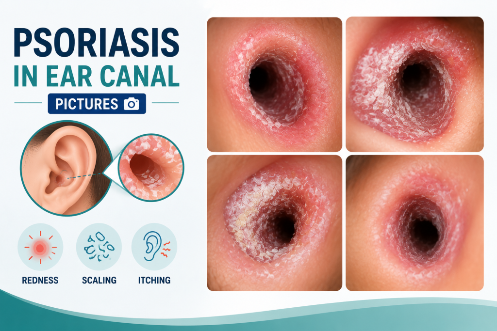
A visual field guide to common patterns, skin-tone differences, mimics, treatment cautions, and hearing-related warning signs.
Searching for psoriasis in ear canal pictures can help you understand the range of possible appearances, but a photo cannot confirm the diagnosis. Psoriasis may affect the outer ear, the folds around the ear, the skin behind it, or the visible portion of the ear canal. Inside the canal, scaling can mix with earwax and become difficult to distinguish from eczema, seborrheic dermatitis, contact irritation, or an infection.
The most useful question is not simply, “Does this look like psoriasis?” It is, “Which visual clues fit psoriasis, which clues point elsewhere, and is there any reason the ear needs to be examined promptly?” This guide answers those questions without encouraging self-treatment inside a sensitive part of the body.
| Quick answer
Ear-canal psoriasis often appears as sharply outlined dry or scaly skin, sometimes with silvery-white, gray, or thicker scale. The surrounding scalp or skin behind the ear may show similar plaques. On deeper skin tones, the inflamed area may look purple, violet-gray, dark brown, or more deeply pigmented rather than bright red. Painful drainage, marked swelling, fever, or sudden hearing change is not a pattern to diagnose from pictures at home. |
Key Takeaways
- Psoriasis in or near the ear usually causes persistent scale, itch, dryness, and a clearly defined patch rather than a single isolated pimple.
- Color varies with skin tone. Redness may be less obvious on brown or Black skin, where plaques can look purple, gray, deep brown, or darker than nearby skin.
- Scale or shed skin can block the ear canal and cause muffled hearing, but hearing loss can have other causes that require an ear examination.
- Do not push cotton swabs, fingernails, tools, creams, or oils into the ear canal unless a clinician has specifically recommended the product and method.
- A dermatologist can confirm the skin diagnosis, while an ear, nose, and throat specialist can safely inspect or clear a blocked canal.
What Do Ear Psoriasis Pictures Usually Show?
The appearance changes according to the exact location, the amount of scale, whether the area has been scratched, and a person’s natural skin color. The following visual map is more useful than relying on one “classic” photo.
| Location in a picture | Typical visual pattern | What the picture cannot tell you |
| At the canal entrance | Dry, flaky, or layered scale around the opening; the skin may appear thickened or cracked. | Whether scale extends deeper, touches the eardrum, or is mixed with impacted wax. |
| Inside the visible canal | Whitish, gray, or silvery scale against inflamed or discolored skin. The canal may look narrowed by debris. | Whether the material is psoriasis scale, eczema, fungus, infection-related debris, or wax. |
| Behind the ear | A sharply bordered plaque, often with cracking in the crease where the ear meets the scalp. | Whether there is bacterial infection in a painful, wet, or crusted fissure. |
| Ear folds and bowl of the ear | Thin or thick scale, redness or discoloration, and possible overlap with greasy yellow scale. | Whether it is plaque psoriasis, inverse psoriasis, or sebopsoriasis. |
| Scalp next to the ear | A larger plaque that crosses the hairline or continues behind the ear. | How much of the scalp is involved or whether treatment is needed throughout the body. |
How can ear psoriasis look different across skin tones?
Many online image collections overrepresent light skin. On lighter skin, an active plaque may look pink or red with white or silvery scale. On medium to deep skin tones, inflammation may appear violet, purple-gray, dark brown, or simply darker than the surrounding skin. Scale can look white, gray, or ashy and may stand out more than the underlying color change. After a flare settles, temporary darkening or lightening can remain, especially on skin of color.
This difference matters because a person may have obvious itch and scaling without the bright redness commonly shown in older medical images. Diagnosis should account for texture, borders, scale, location, symptoms, and other psoriasis patches, not color alone.
A Five-Clue Picture Check
- Border: Psoriasis often has a more clearly defined edge than simple dryness. The patch may end abruptly where normal skin begins.
- Scale: Dry white, gray, or silvery scale supports psoriasis. Greasy yellow scale suggests seborrheic dermatitis or sebopsoriasis, although overlap is possible.
- Distribution: A matching plaque on the scalp, hairline, elbows, knees, or behind the other ear increases suspicion for psoriasis.
- Surface damage: Scratching can produce cracks, pinpoint bleeding, crusting, or raw areas. These changes make pictures harder to interpret and increase infection risk.
- Ear symptoms: Muffled hearing can occur when scale blocks the canal. Strong pain, pus-like drainage, fever, or rapidly increasing swelling points away from an uncomplicated plaque and needs medical review.
| Photo limitation
A smartphone picture usually cannot show the entire ear canal or eardrum. Lighting changes the apparent color, and automatic camera processing can exaggerate redness or wash out purple and brown tones. An otoscope examination is more reliable when symptoms are inside the canal. |
What Can Look Like Psoriasis in Ear Canal Pictures?
Several common conditions create overlapping itch, scale, and discoloration. A comparison can narrow the possibilities, but it should not be used to place medication deep in the ear.
| Possible look-alike | Clues in a picture | Clues from symptoms | Why an exam may matter |
| Ear eczema | Dry, cracked, inflamed skin; may be less sharply bordered. | Often very itchy; may be linked with allergies or eczema elsewhere. | Treatment differs, and the canal may be fragile or infected. |
| Seborrheic dermatitis | Greasy or yellowish scale, commonly around the scalp and outer ear. | Itch and flaking may fluctuate with oily areas of the face and scalp. | It can overlap with psoriasis as sebopsoriasis. |
| Contact dermatitis | Inflammation where earbuds, hearing aids, hair products, jewelry, or drops touch the skin. | Burning or itching after a new product or repeated exposure. | The trigger must be identified and the eardrum status may affect drop selection. |
| Otitis externa | Swollen canal, wet debris, or drainage rather than only dry scale. | Pain when the outer ear is moved, tenderness, discharge, or recent swimming. | An infection can worsen and usually needs targeted treatment. |
| Fungal otitis externa | Fluffy, speckled, or dark debris may be visible, but photos vary. | Persistent itch, fullness, drainage, or recent antibiotic ear drops. | Microscopic or otoscopic examination helps distinguish fungus from scale. |
| Impacted earwax | Brown, tan, orange, or dark material filling the canal. | Fullness, muffled hearing, or no skin symptoms at all. | Home tools can push wax deeper or injure the canal and eardrum. |
Can a Doctor Diagnose Ear Psoriasis From a Picture?
A clear image may help with triage, especially when the affected skin is on the outer ear or behind it. Diagnosis usually depends on a skin examination, medical history, and a search for psoriasis elsewhere. When symptoms are inside the canal, a clinician may use an otoscope to inspect the canal and eardrum. A biopsy is uncommon but may be considered when the diagnosis remains uncertain.
A dermatologist is often the best starting point when there are plaques on the scalp or body. An ear, nose, and throat specialist may be needed when scale or wax blocks the canal, hearing has changed, the diagnosis is unclear, or the ear requires safe cleaning. The National Psoriasis Foundation specifically advises against putting fingers or cotton swabs into the canal and recommends ENT help for blockage.
What Does Treatment Look Like in 2026?
Treatment is chosen according to location. A product that is appropriate behind the ear may not be appropriate deep in the canal. The goal is to calm inflammation, reduce scale, prevent blockage, and control psoriasis elsewhere so the ear is not treated in isolation.
- Treat the visible skin carefully: A clinician may recommend a topical steroid or a nonsteroidal topical medicine for limited disease. Potency, formulation, frequency, and duration matter because ear skin is thin and sensitive.
- Address scale without digging: Loose scale on the outer ear can be handled gently according to a care plan. Material blocking the canal should be removed by a healthcare professional rather than with swabs, picks, candles, or suction gadgets.
- Control broader psoriasis: If the ear is part of more extensive or difficult psoriasis, phototherapy, oral treatment, or biologic treatment may be considered. Better overall disease control can reduce ear flares.
- Check products and devices: Earbuds, hearing aids, hair dye, fragrance, shampoo, and ear drops can add friction or contact irritation. Cleaning devices as directed and identifying triggers may reduce overlapping inflammation.
- Recheck hearing: Hearing that remains muffled after the visible flare improves deserves an ear examination and, when indicated, formal hearing testing.
What should you avoid putting in the ear canal?
- Cotton swabs, fingernails, hairpins, curettes, or other objects
- Over-the-counter psoriasis cream unless a clinician says it is safe for that exact location
- Hydrogen peroxide, alcohol, essential oils, vinegar mixtures, or home remedies when the eardrum has not been examined
- Ear candles, which do not safely remove blockage and can cause burns or injury
- Repeated scratching or picking, which can worsen psoriasis through skin trauma and can introduce infection
When Is an Ear Picture Not Enough?
| Arrange medical care | Seek urgent or emergency evaluation |
|
· New or persistent scaling inside the canal · Muffled hearing, fullness, or recurrent blockage · Pain, swelling, oozing, crusting, or bad odor · A rash that does not improve with the prescribed plan · Uncertainty about psoriasis versus eczema or infection |
· Sudden hearing loss in one or both ears · Severe ear pain with fever or rapidly spreading redness · New facial weakness, severe dizziness, fainting, or neurologic symptoms · Significant bleeding or suspected eardrum injury · Severe symptoms in a person with diabetes or a weakened immune system |
Sudden unexplained hearing loss is time-sensitive. The National Institute on Deafness and Other Communication Disorders advises treating sudden sensorineural hearing loss as a medical emergency rather than assuming the canal is merely blocked.
How Can You Take a Useful Photo for a Telehealth Visit?
Photograph only what is safely visible. Do not insert the phone, a camera attachment, or another device into the canal. A useful set usually includes:
- One image showing the entire ear and nearby scalp
- One closer image of the affected outer-ear skin in indirect daylight
- A matching image of the other ear for comparison
- A photo of psoriasis elsewhere, if present
- A brief note stating when the rash began, whether it itches or hurts, and whether hearing changed
Avoid filters, flash glare, and heavy image sharpening. A photo can document change over time, but it should not delay an in-person ear examination when there is drainage, severe pain, canal blockage, or hearing loss.
Questions People Ask After Comparing Pictures
Can ear psoriasis look like earwax?
Yes. White or gray scale may mix with brown wax and form a plug. Color alone cannot reliably separate the two. An otoscope examination can show whether the canal is blocked and whether the eardrum is visible.
Does ear psoriasis always affect both ears?
No. It may affect one ear, both ears, or alternate sides during different flares. One-sided disease does not rule it out, but one-sided pain or drainage increases the need to exclude infection or another local cause.
Is psoriasis inside the ear contagious?
No. Psoriasis is an immune-mediated disease and cannot be spread by touching the rash. A secondary infection can occur in broken skin, however, and that requires separate treatment.
Can it cause permanent hearing loss?
A plug of scale or wax can cause temporary conductive hearing loss that may improve after safe removal. Research has also found associations between psoriasis and other types of hearing or balance problems, but the clinical meaning varies. Persistent or sudden hearing changes should be evaluated rather than attributed to skin disease.
Why does it keep returning behind the ear?
The crease is exposed to moisture, friction, hair products, and scratching. Psoriasis is also chronic, so plaques can recur even after successful treatment. A maintenance plan and control of nearby scalp psoriasis may reduce repeated flares.
Can I use dandruff shampoo around my ears?
A medicated scalp product may help nearby scalp psoriasis or seborrheic dermatitis, but it can irritate thin ear skin and should not enter the canal. Follow the product label and a dermatologist’s instructions for the ear area.
| The practical takeaway
Pictures can help you recognize a scaly pattern, compare both ears, and notice changes across skin tones. They cannot show the full canal, prove the cause, or rule out infection. The safest path is a dermatology assessment for the skin condition and an ENT examination when debris, pain, drainage, dizziness, or hearing change involves the canal. |
Authoritative Sources and Further Reading
- National Psoriasis Foundation: Psoriasis in the Ears (medically reviewed March 23, 2026)
- Cleveland Clinic: Psoriasis in the Ears
- NIDCD: Sudden Deafness
Medical disclaimer: This article is for general educational purposes and does not diagnose a skin or ear condition. Seek care from a qualified U.S. healthcare professional for individualized evaluation and treatment.